Home > Product > Antibody > Rabbit Anti-Cathepsin B antibody
Amyloid precursor protein secretase; APP secretase; APPS; Cathepsin B; CB; Cathepsin B1; CathepsinB; CPSB; CTSB; Cysteine protease; Preprocathepsin B; Cathepsin B heavy chain; Cathepsin B light chain; CATB_HUMAN.
Cat:
SL300R
Species Reactivity:
Human,Mouse,Rat,
Immunogen:
KLH conjugated synthetic peptide derived from human Cathepsin B heavy chain:251-339/339
Format:
Liquid
Storage instructions:
Shipped at 4℃. Store at -20 °C for one year. Avoid repeated freeze/thaw cycles.
Concentration:
1mg/ml
Clonality:
Polyclonal
Isotype:
IgG
Applications:
WB=1:500-2000ELISA=1:5000-10000IHC-P=1:100-500IHC-F=1:100-500Flow-Cyt=1µg/TestICC=1:100IF=1:100-500(Paraffin sections need to do antigen repair)not yet tested in other applications.optimal dilutions/concentrations should be determined by the end user.
Host:
Rabbit
Product Overview:
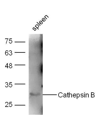
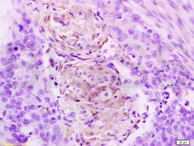
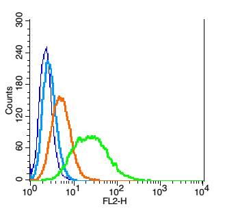
Sample: Spleen (Mouse) Lysate at 40 ugPrimary: Anti-Cathepsin B (SL300R) at 1/300 dilutionSecondary: IRDye800CW Goat Anti-Rabbit IgG at 1/20000 dilutionPredicted band size: 46/28 kDObserved band size: 30 kDTissue/cell: Human esophageal carcinoma; 4% Paraformaldehyde-fixed and paraffin-embedded; Antigen retrieval: citrate buffer ( 0.01M, pH 6.0 ), Boiling bathing for 15min; Block endogenous peroxidase by 3% Hydrogen peroxide for 30min; Blocking buffer (normal goat serum,SLC0005) at 37℃ for 20 min; Incubation: Anti-Cathepsin B Polyclonal Antibody, Unconjugated(SL300R) 1:200, overnight at 4°C, followed by conjugation to the secondary antibody(SP-0023) and DAB(SLC0010) stainingBlank control: RSC96(blue). Primary Antibody:Rabbit Anti- Cathepsin B antibody(SL300R), Dilution: 1μg in 100 μL 1X PBS containing 0.5% BSA; Isotype Control Antibody: Rabbit IgG(orange) ,used under the same conditions ); Secondary Antibody: Goat anti-rabbit IgG-PE(white blue), Dilution: 1:200 in 1 X PBS containing 0.5% BSA.ProtocolThe cells were fixed with 2% paraformaldehyde (10 min) . Antibody (SL300R, 5μg /1x10^6 cells) were incubated for 30 min on the ice, followed by 1 X PBS containing 0.5% BSA + 1 0% goat serum (15 min) to block non-specific protein-protein interactions. Then the Goat Anti-rabbit IgG/PE antibody was added into the blocking buffer mentioned above to react with the primary antibody of SL300R at 1/200 dilution for 30 min on ice. Acquisition of 20,000 events was performed.
More
Unit:
Price: $
Product PDFs
Datasheet:


The protein encoded by this gene is a lysosomal cysteine proteinase composed of a dimer of disulfide-linked heavy and light chains, both produced from a single protein precursor. It is also known as amyloid precursor protein secretase and is involved in the proteolytic processing of amyloid precursor protein (APP). Incomplete proteolytic processing of APP has been suggested to be a causative factor in Alzheimer disease, the most common cause of dementia. Overexpression of the encoded protein, which is a member of the peptidase C1 family, has been associated with esophageal adenocarcinoma and other tumors. At least five transcript variants encoding the same protein have been found for this gene. [provided by RefSeq, Jul 2008]

Function:
Thiol protease which is believed to participate in intracellular degradation and turnover of proteins. Has also been implicated in tumor invasion and metastasis.

Subunit:
Dimer of a heavy chain and a light chain cross-linked by a disulfide bond. Interacts with SRPX2.

Subcellular Location:
Lysosome. Melanosome. Note=Identified by mass spectrometry in melanosome fractions from stage I to stage IV.

Similarity:
Belongs to the peptidase C1 family.

SWISS:
P10605

Gene ID:
1508

Database links:

Entrez Gene: 1508 Human

Entrez Gene: 13030 Mouse

Entrez Gene: 64529 Rat

Omim: 116810 Human

SwissProt: P07858 Human

SwissProt: P10605 Mouse

SwissProt: P00787 Rat

Unigene: 520898 Human

Unigene: 236553 Mouse

Unigene: 472420 Mouse

Unigene: 100909 Rat


合成与降解(Synthesis and Degradation)
组织蛋白酶B(CB) 参与肿瘤的侵袭转移过程。可促进肿瘤血管的增生,增强肿瘤细胞的运动能力.组织蛋白酶B转录以及表达水平的高低与肿瘤预后呈正相关。
cathepsin B属于半胱氨酸蛋白酶,与人体的多种生理、病理过程密切相关.组织蛋白酶B (cathepsin B,CB)主要存在于溶酶体中, 发挥其生理状态下的蛋白水解作用.但在某些特定条件下, CB在胞外间质中有时也可以检测到.
目前,有学者将CB用于阿尔茨海默病(AD)方面的研究。 Picture

Sample:
Spleen (Mouse) Lysate at 40 ug
Primary: Anti-Cathepsin B (SL300R) at 1/300 dilution
Secondary: IRDye800CW Goat Anti-Rabbit IgG at 1/20000 dilution
Predicted band size: 46/28 kD
Observed band size: 30 kD
Tissue/cell: Human esophageal carcinoma; 4% Paraformaldehyde-fixed and paraffin-embedded;
Antigen retrieval: citrate buffer ( 0.01M, pH 6.0 ), Boiling bathing for 15min; Block endogenous peroxidase by 3% Hydrogen peroxide for 30min; Blocking buffer (normal goat serum,SLC0005) at 37℃ for 20 min;
Incubation: Anti-Cathepsin B Polyclonal Antibody, Unconjugated(SL300R) 1:200, overnight at 4°C, followed by conjugation to the secondary antibody(SP-0023) and DAB(SLC0010) staining
Blank control: RSC96(blue).
Primary Antibody:Rabbit Anti- Cathepsin B antibody(SL300R), Dilution: 1μg in 100 μL 1X PBS containing 0.5% BSA;
Isotype Control Antibody: Rabbit IgG(orange) ,used under the same conditions );
Secondary Antibody: Goat anti-rabbit IgG-PE(white blue), Dilution: 1:200 in 1 X PBS containing 0.5% BSA.
Protocol
The cells were fixed with 2% paraformaldehyde (10 min) . Antibody (SL300R, 5μg /1x10^6 cells) were incubated for 30 min on the ice, followed by 1 X PBS containing 0.5% BSA + 1 0% goat serum (15 min) to block non-specific protein-protein interactions. Then the Goat Anti-rabbit IgG/PE antibody was added into the blocking buffer mentioned above to react with the primary antibody of SL300R at 1/200 dilution for 30 min on ice. Acquisition of 20,000 events was performed.
Product Feedback Wall
Message :
Your Email :
Copyright © 2007-2018 Sunlong Medical All Rights Reserved.