VCAM1 is important in cell-cell recognition. Appears to function in leukocyte-endothelial cell adhesion. Interacts with the integrins alpha4 beta1 (beta 1 integrin VLA4) and alpha4 beta7 on leukocytes, and mediates both adhesion and signal transduction. The VCAM1/VLA4 interaction may play a pathophysiologic role both in immune responses and in leukocyte emigration to sites of inflammation. VCAM1 is also expressed by several non endothelial cell types including some macrophages, follicular dendritic cells and bone marrow, stromal cells.
Function:
Important in cell-cell recognition. Appears to function in leukocyte-endothelial cell adhesion. Interacts with the beta-1 integrin VLA4 on leukocytes, and mediates both adhesion and signal transduction. The VCAM1/VLA4 interaction may play a pathophysiologic role both in immune responses and in leukocyte emigration to sites of inflammation.
Subunit:
Binds to ECMSLVD capsid proteins and acts as a receptor for this virus.
Subcellular Location:
Isoform 1: Cell membrane; Single-pass type I membrane protein. Isoform 2: Cell membrane; Lipid-anchor, GPI-anchor.
Tissue Specificity:
Expressed on inflamed vascular endothelium, as well as on macrophage-like and dendritic cell types in both normal and inflamed tissue. Expressed in the bone marrow.
Similarity:
Contains 7 Ig-like C2-type (immunoglobulin-like) domains.
SWISS:
P29533
Gene ID:
22329
Database links:
Entrez Gene: 7412 Human
Entrez Gene: 22329 Mouse
Entrez Gene: 25361 Rat
GenBank: NP_001069.1 Human
GenBank: NP_001186763.1 Human
Omim: 192225 Human
SwissProt: P1964 Human
SwissProt: P29533 Mouse
VCAM 1(CD106)是一种膜相关蛋白,分布于血管内皮及其周围的淋巴细胞、单核细胞、嗜酸性粒细胞和中性粒细胞。Anti-VCAM-1主要用于各种组织中血管内皮的检测。
| Picture |
Sample:
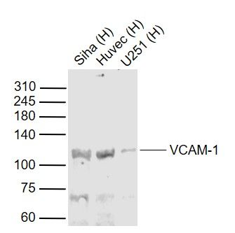
Lane 1: Siha (Human) Cell Lysate at 30 ug
Lane 2: Huvec (Human) Cell Lysate at 30 ug
Lane 3: U251 (Human) Cell Lysate at 30 ug
Primary: Anti-VCAM-1 (SL0396R) at 1/1000 dilution
Secondary: IRDye800CW Goat Anti-Rabbit IgG at 1/20000 dilution
Predicted band size: 110 kD
Observed band size: 110 kD
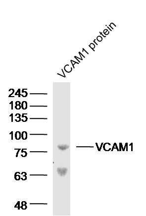
Sample: VCAM1 protein (Human) at 100 ng
Primary: Anti-VCAM-1 (SL0396R) at 1/300 dilution
Secondary: IRDye800CW Goat Anti-Rabbit IgG at 1/20000 dilution
Predicted band size: 81 kD
Observed band size: 81 kD
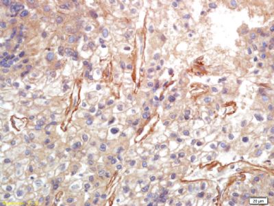
Tissue/cell: human lung carcinoma; 4% Paraformaldehyde-fixed and paraffin-embedded;
Antigen retrieval: citrate buffer ( 0.01M, pH 6.0 ), Boiling bathing for 15min; Block endogenous peroxidase by 3% Hydrogen peroxide for 30min; Blocking buffer (normal goat serum,SLC0005) at 37℃ for 20 min;
Incubation: Anti-VCAM-1 Polyclonal Antibody, Unconjugated(SL0396R) 1:200, overnight at 4°C, followed by conjugation to the secondary antibody(SP-0023) and DAB(SLC0010) staining
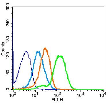
Blank control: Mouse Spleen(blue).
Primary Antibody:Rabbit Anti-VCAM-1 antibody (SL0396R,Green); Dilution: 1μg in 100 μL 1X PBS containing 0.5% BSA;
Isotype Control Antibody: Rabbit IgG(orange) ,used under the same conditions;
Secondary Antibody: Goat anti-rabbit IgG-FITC(white blue), Dilution: 1:200 in 1 X PBS containing 0.5% BSA.
Protocol
The cells were fixed with 2% paraformaldehyde for 10 min at 37℃. Primary antibody (SL0396R, 1μg /8x10^5 cells) were incubated for 30 min at room temperature, followed by 1 X PBS containing 0.5% BSA + 1 0% goat serum (1 hour) to block non-specific protein-protein interactions. Then the Goat Anti-rabbit IgG/FITC antibody was added into the blocking buffer mentioned above to react with the primary antibody at 1/200 dilution for 40 min at room temperature. Acquisition of 20,000 events was performed.
|
|
|