Home > Product > Antibody > Rabbit Anti-Collagen III antibody
COL 3A1; COL3A1; Collagen alpha 1(III) chain; Collagen III alpha 1 chain precursor; Collagen III alpha 1 polypeptide; Collagen type III alpha 1 (Ehlers Danlos syndrome type IV autosomal dominant); Collagen type III alpha 1; Collagen type III alpha; EDS4A;
Cat:
SL0948R
Species Reactivity:
Human,Rat,(predicted: Mouse,Cow,)
Immunogen:
KLH conjugated synthetic peptide derived from human Collagen alpha 1(III) chain:801-900/1466
Format:
Liquid
Storage instructions:
Shipped at 4℃. Store at -20 °C for one year. Avoid repeated freeze/thaw cycles.
Concentration:
1mg/ml
Clonality:
Polyclonal
Isotype:
IgG
Applications:
WB=1:500-2000ELISA=1:5000-10000IHC-P=1:100-500IHC-F=1:100-500IF=1:100-500(Paraffin sections need to do antigen repair)not yet tested in other applications.optimal dilutions/concentrations should be determined by the end user.
Host:
Rabbit
Product Overview:
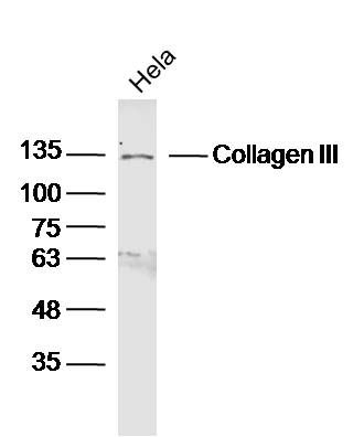
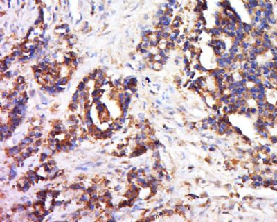
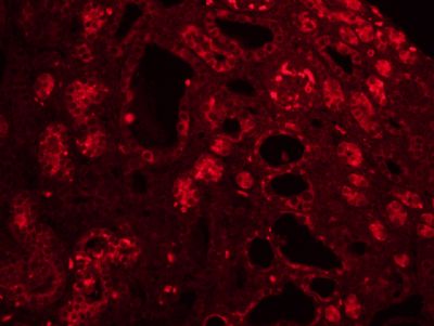
Sample: Hela Cell (Human) Lysate at 40 ugPrimary: Anti- Collagen III (SL0948R) at 1/300 dilutionSecondary: IRDye800CW Goat Anti-Rabbit IgG at 1/20000 dilutionPredicted band size: 117 kDObserved band size: 130 kDTissue/cell: human colon cancer; 4% Paraformaldehyde-fixed and paraffin-embedded; Antigen retrieval: citrate buffer ( 0.01M, pH 6.0 ), Boiling bathing for 15min; Block endogenous peroxidase by 3% Hydrogen peroxide for 30min; Blocking buffer (normal goat serum,SLC0005) at 37℃ for 20 min; Incubation: Anti-Collagen III Polyclonal Antibody, Unconjugated(SL0948R) 1:500, overnight at 4°C, followed by conjugation to the secondary antibody(SP-0023) and DAB(SLC0010) stainingTissue/cell: rat kidney tissue;4% Paraformaldehyde-fixed and paraffin-embedded; Antigen retrieval: citrate buffer ( 0.01M, pH 6.0 ), Boiling bathing for 15min; Blocking buffer (normal goat serum,SLC0005) at 37℃ for 20 min; Incubation: Anti-Collagen III Polyclonal Antibody, Unconjugated(SL0948R) 1:200, overnight at 4°C; The secondary antibody was Goat Anti-Rabbit IgG, Cy3 conjugated(SL0295G-Cy3)used at 1:200 dilution for 40 minutes at 37°C.
More
Unit:
Price: $
Product PDFs
Datasheet:


The extensive family of COL gene products (collagens) is composed of several chain types, including fibril-forming interstitial collagens (types I, II, III and V) and basement membrane collagens (type IV), each type containing multiple isoforms. Collagens are fibrous, extracellular matrix proteins with high tensile strength and are the major components of connective tissue, such as tendons and cartilage. All collagens contain a triple helix domain and frequently show lateral self-association in order to form complex connective tissues. Several collagens also play a role in cell adhesion, important for maintaining normal tissue architecture and function.

This gene encodes the pro-alpha1 chains of type III collagen, a fibrillar collagen that is found in extensible connective tissues such as skin, lung, uterus, intestine and the vascular system, frequently in association with type I collagen. Mutations in this gene are associated with Ehlers-Danlos syndrome types IV, and with aortic and arterial aneurysms. Two transcripts, resulting from the use of alternate polyadenylation signals, have been identified for this gene.

Function:
Collagen type III occurs in most soft connective tissues along with type I collagen.

Subunit:
Trimers of identical alpha 1(III) chains. The chains are linked to each other by interchain disulfide bonds. Trimers are also cross-linked via hydroxylysines.

Subcellular Location:
Secreted, extracellular space, extracellular matrix.

Post-translational modifications:
Proline residues at the third position of the tripeptide repeating unit (G-X-Y) are hydroxylated in some or all of the chains.
O-linked glycan consists of a Glc-Gal disaccharide bound to the oxygen atom of a post-translationally added hydroxyl group.

DISEASE:
Defects in COL3A1 are a cause of Ehlers-Danlos syndrome type 3 (EDS3) [MIM:130020]; also known as benign hypermobility syndrome. EDS is a connective tissue disorder characterized by hyperextensible skin, atrophic cutaneous scars due to tissue fragility and joint hyperlaxity. EDS3 is a form of Ehlers-Danlos syndrome characterized by marked joint hyperextensibility without skeletal deformity.
Defects in COL3A1 are the cause of Ehlers-Danlos syndrome type 4 (EDS4) [MIM:130050]. EDS is a connective tissue disorder characterized by hyperextensible skin, atrophic cutaneous scars due to tissue fragility and joint hyperlaxity. EDS4 is the most severe form of the disease. It is characterized by the joint and dermal manifestations as in other forms of the syndrome, characteristic facial features (acrogeria) in most patients, and by proneness to spontaneous rupture of bowel and large arteries. The vascular complications may affect all anatomical areas.
Defects in COL3A1 are a cause of susceptibility to aortic aneurysm abdominal (AAA) [MIM:100070]. AAA is a common multifactorial disorder characterized by permanent dilation of the abdominal aorta, usually due to degenerative changes in the aortic wall. Histologically, AAA is characterized by signs of chronic inflammation, destructive remodeling of the extracellular matrix, and depletion of vascular smooth muscle cells.

Similarity:
Belongs to the fibrillar collagen family.
Contains 1 fibrillar collagen NC1 domain.
Contains 1 VWFC domain.

SWISS:
P02461

Gene ID:
1281

Database links:

Entrez Gene: 510833 Cow

Entrez Gene: 1281 Human

Entrez Gene: 12825 Mouse

Entrez Gene: 16832 Rat

Omim: 12036 Human

SwissProt: P04258 Cow

SwissProt: P02461 Human

SwissProt: P08121 Mouse

SwissProt: P13941 Rat

Unigene: 443625 Human

Unigene: 249555 Mouse

Unigene: 3247 Rat


Picture

Sample:
Hela Cell (Human) Lysate at 40 ug
Primary: Anti- Collagen III (SL0948R) at 1/300 dilution
Secondary: IRDye800CW Goat Anti-Rabbit IgG at 1/20000 dilution
Predicted band size: 117 kD
Observed band size: 130 kD
Tissue/cell: human colon cancer; 4% Paraformaldehyde-fixed and paraffin-embedded;
Antigen retrieval: citrate buffer ( 0.01M, pH 6.0 ), Boiling bathing for 15min; Block endogenous peroxidase by 3% Hydrogen peroxide for 30min; Blocking buffer (normal goat serum,SLC0005) at 37℃ for 20 min;
Incubation: Anti-Collagen III Polyclonal Antibody, Unconjugated(SL0948R) 1:500, overnight at 4°C, followed by conjugation to the secondary antibody(SP-0023) and DAB(SLC0010) staining
Tissue/cell: rat kidney tissue;4% Paraformaldehyde-fixed and paraffin-embedded;
Antigen retrieval: citrate buffer ( 0.01M, pH 6.0 ), Boiling bathing for 15min; Blocking buffer (normal goat serum,SLC0005) at 37℃ for 20 min;
Incubation: Anti-Collagen III Polyclonal Antibody, Unconjugated(SL0948R) 1:200, overnight at 4°C; The secondary antibody was Goat Anti-Rabbit IgG, Cy3 conjugated(SL0295G-Cy3)used at 1:200 dilution for 40 minutes at 37°C.
Product Feedback Wall
Message :
Your Email :
Copyright © 2007-2018 Sunlong Medical All Rights Reserved.