Hexokinases phosphorylate glucose to produce glucose 6 phosphate, thus committing glucose to the glycolytic pathway. Alternative splicing of this gene results in three tissue specific forms of glucokinase, one found in pancreatic islet beta cells and two found in liver. The protein localizes to the outer membrane of mitochondria. In contrast to other forms of hexokinase, this enzyme is not inhibited by its product glucose 6 phosphate but remains active while glucose is abundant. Mutations in this gene have been associated with non insulin dependent diabetes mellitus, also called maturity onset diabetes of the young, type 2; mutations have also been associated with persistent hyperinsulinemic hypoglycemia of infancy (PHHI).
Function:
Catalyzes the initial step in utilization of glucose by the beta-cell and liver at physiological glucose concentration. Glucokinase has a high Km for glucose, and so it is effective only when glucose is abundant. The role of GCK is to provide G6P for the synthesis of glycogen. Pancreatic glucokinase plays an important role in modulating insulin secretion. Hepatic glucokinase helps to facilitate the uptake and conversion of glucose by acting as an insulin-sensitive determinant of hepatic glucose usage.
Subunit:
Monomer.
Tissue Specificity:
Isoform 1 is expressed in pancreas. Isoform 2 and isoform 3 is expressed in liver.
DISEASE:
Defects in GCK are the cause of maturity-onset diabetes of the young type 2 (MODY2) [MIM:125851]; also shortened MODY-2. MODY is a form of diabetes that is characterized by an autosomal dominant mode of inheritance, onset in childhood or early adulthood (usually before 25 years of age), a primary defect in insulin secretion and frequent insulin-independence at the beginning of the disease.
Defects in GCK are the cause of familial hyperinsulinemic hypoglycemia type 3 (HHF3) [MIM:602485]; also known as persistent hyperinsulinemic hypoglycemia of infancy (PHHI) or congenital hyperinsulinism. HHF is the most common cause of persistent hypoglycemia in infancy. Unless early and aggressive intervention is undertaken, brain damage from recurrent episodes of hypoglycemia may occur.
Similarity:
Belongs to the hexokinase family.
SWISS:
P35557
Gene ID:
2645
Database links:
Entrez Gene: 2645 Human
Entrez Gene: 103988 Mouse
Entrez Gene: 24385 Rat
Omim: 27679 Human
SwissProt: P35557 Human
SwissProt: P52792 Mouse
SwissProt: P17712 Rat
Unigene: 1270 Human
Unigene: 220358 Mouse
Unigene: 10447 Rat
| Picture |
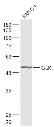
Sample:
Panc-1(Human) Cell Lysate at 30 ug
Primary: Anti-GLK (SL1796R) at 1/1000 dilution
Secondary: IRDye800CW Goat Anti-Rabbit IgG at 1/20000 dilution
Predicted band size: 51 kD
Observed band size: 51 kD
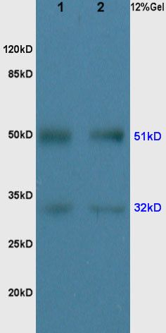
Protein: rat liver lysates, 30ug;
Primary: Anti-GLK(SL1796R) at 1:200;
Secondary: HRP conjugated Goat Anti-Rabbit IgG(SL0295G-HRP) at 1: 3000;
ECL excitated the fluorescence;
Predicted band size : 51kD
Observed band size : 51kD
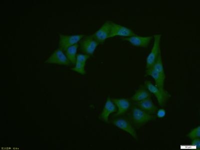
Hela cell; 4% Paraformaldehyde-fixed; Triton X-100 at room temperature for 20 min; Blocking buffer (normal goat serum, SLC0005) at 37°C for 20 min; Antibody incubation with (GLK) polyclonal Antibody, Unconjugated (SL1796R) 1:100, 90 minutes at 37°C; followed by a conjugated Goat Anti-Rabbit IgG antibody at 37°C for 90 minutes, DAPI (blue, C02-04002) was used to stain the cell nuclei.
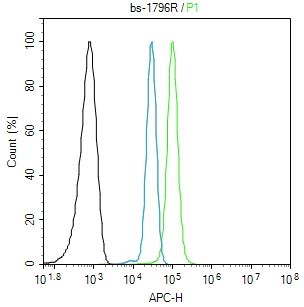
Blank control: A431.
Primary Antibody (green line): Rabbit Anti-GLK antibody (SL1796R)
Dilution: 1μg /10^6 cells;
Isotype Control Antibody (orange line): Rabbit IgG .
Secondary Antibody: Goat anti-rabbit IgG-AF647
Dilution: 1μg /test.
Protocol
The cells were fixed with 4% PFA (10min at room temperature)and then permeabilized with 0.1% PBST for 20 min at room temperature. The cells were then incubated in 5%BSA to block non-specific protein-protein interactions for 30 min at room temperature .Cells stained with Primary Antibody for 30 min at room temperature. The secondary antibody used for 40 min at room temperature. Acquisition of 20,000 events was performed.
|
|
|