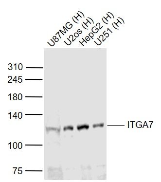
Sample:
Lane 1: U87MG (Human) Cell Lysate at 30 ug
Lane 2: U2os (Human) Cell Lysate at 30 ug
Lane 3: HepG2 (Human) Cell Lysate at 30 ug
Lane 4: U251 (Human) Cell Lysate at 30 ug
Primary: Anti-ITGA7 (SL1816R) at 1/1000 dilution
Secondary: IRDye800CW Goat Anti-Rabbit IgG at 1/20000 dilution
Predicted band size: 120 kD
Observed band size: 120 kD
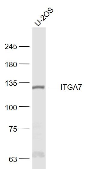
Sample:
U-2OS(Human) Cell Lysate at 30 ug
Primary: Anti-ITGA7 (SL1816R) at 1/1000 dilution
Secondary: IRDye800CW Goat Anti-Rabbit IgG at 1/20000 dilution
Predicted band size: 70/101/125 kD
Observed band size: 125 kD
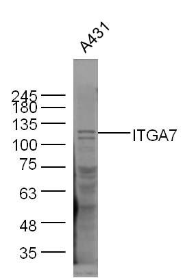
Sample:
A431(Human) Cell Lysate at 30 ug
Primary: Anti-ITGA7 (SL 1816R) at 1/300 dilution
Secondary: IRDye800CW Goat Anti-Rabbit IgG at 1/20000 dilution
Predicted band size: 125 kD
Observed band size: 125 kD
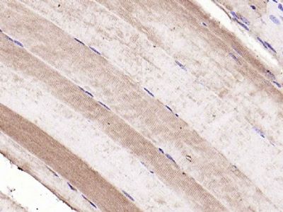
Paraformaldehyde-fixed, paraffin embedded (mouse skeletal muscle); Antigen retrieval by boiling in sodium citrate buffer (pH6.0) for 15min; Block endogenous peroxidase by 3% hydrogen peroxide for 20 minutes; Blocking buffer (normal goat serum) at 37°C for 30min; Antibody incubation with (ITGA7) Polyclonal Antibody, Unconjugated (SL1816R) at 1:200 overnight at 4°C, followed by operating according to SP Kit(Rabbit) (sp-0023) instructionsand DAB staining.
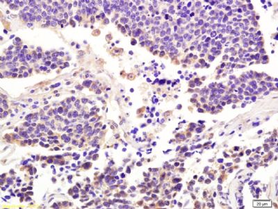
Paraformaldehyde-fixed, paraffin embedded (human lung carcinoma) ; Antigen retrieval by boiling in sodium citrate buffer (pH6) for 15min; Block endogenous peroxidase by 3% hydrogen peroxide for 30 minutes; Blocking buffer (normal goat serum) at 37°C for 20min; Antibody incubation with Integrin alpha 7 Polyclonal Antibody, Unconjugated (SL1816R) at 1:200 overnight at 4°C, followed by a conjugated secondary and DAB staining.
|