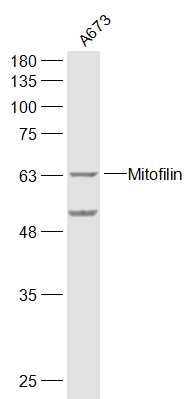
Sample:
A673(Human) Cell Lysate at 30 ug
Primary: Anti-Mitofilin (SL1824R) at 1/300 dilution
Secondary: IRDye800CW Goat Anti-Rabbit IgG at 1/20000 dilution
Predicted band size: 68 kD
Observed band size: 68 kD
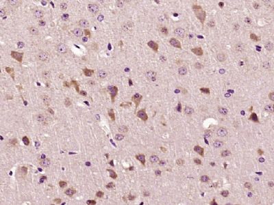
Paraformaldehyde-fixed, paraffin embedded (mouse brain tissue); Antigen retrieval by boiling in sodium citrate buffer (pH6.0) for 15min; Block endogenous peroxidase by 3% hydrogen peroxide for 20 minutes; Blocking buffer (normal goat serum) at 37°C for 30min; Antibody incubation with (Mitofilin) Polyclonal Antibody, Unconjugated (SL1824R) at 1:400 overnight at 4°C, followed by operating according to SP Kit(Rabbit) (sp-0023) instructionsand DAB staining.
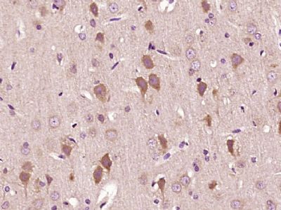
Paraformaldehyde-fixed, paraffin embedded (rat brain tissue); Antigen retrieval by boiling in sodium citrate buffer (pH6.0) for 15min; Block endogenous peroxidase by 3% hydrogen peroxide for 20 minutes; Blocking buffer (normal goat serum) at 37°C for 30min; Antibody incubation with (Mitofilin) Polyclonal Antibody, Unconjugated (SL1824R) at 1:400 overnight at 4°C, followed by operating according to SP Kit(Rabbit) (sp-0023) instructionsand DAB staining.
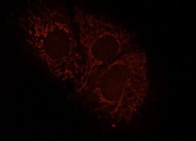
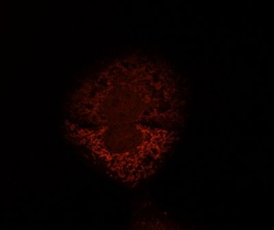
Human HepG2 cells fixed with 3.7% Formaldehyde in medium, blocking performed with 10% NGS in 1x PBS buffer + 0.1% Tween20. Incubated with Anti-Mitofilin/IMMT Polyclonal Antibody (SL1824R) at a 1:10 dilution for 1hr at RT, followed by Anti-Rabbit secondary, Cy3 conjugate at a 1:300 dilution incubated at RT for 1hr. This image was kindly provided by an end-user.
Human HepG2 cells fixed with 3.7% Formaldehyde in medium, blocking performed with 10% NGS in 1x PBS buffer + 0.1% Tween20. Incubated with Anti-Mitofilin/IMMT Polyclonal Antibody (SL1824R) at a 1:10 dilution for 1hr at RT, followed by Anti-Rabbit secondary, Cy3 conjugate at a 1:300 dilution incubated at RT for 1hr. This image was kindly provided by an end-user.
|