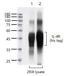
Sample:
Lane 1: IL-4R-His protein (1-232) Overexpression 293T (Human) Cell Lysate at 4 ug
Lane 2: IL-4R-His protein (1-232) Overexpression 293T (Human) Cell Lysate at 4 ug
Primary:
Lane 1: Anti-IL-4R (SL22901R) at 1/1000 dilution
Lane 2: Anti-His tag (SLM33004M) at 1/1000 dilution
Secondary:
Lane 1: IRDye800CW Goat Anti-Rabbit IgG at 1/20000 dilution
Lane 2: IRDye800CW Goat Anti-Mouse IgG at 1/20000 dilution
Predicted band size: 43-48 kD
Observed band size: 43-48 kD
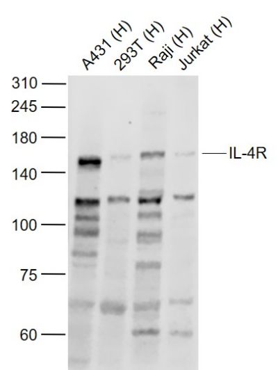
Sample:
Lane 1: A431 (Human) Cell Lysate at 30 ug
Lane 2: 293T (Human) Cell Lysate at 30 ug
Lane 3: Raji (Human) Cell Lysate at 30 ug
Lane 4: Jurkat (Human) Cell Lysate at 30 ug
Primary: Anti-IL-4R (SL22901R) at 1/1000 dilution
Secondary: IRDye800CW Goat Anti-Rabbit IgG at 1/20000 dilution
Predicted band size: 140-160 kD
Observed band size: 150 kD
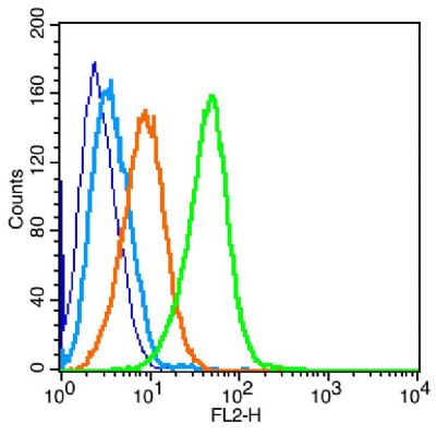
Blank control: Raji (blue).
Primary Antibody:Rabbit Anti-IL-4R antibody(SL2458R), Dilution: 1μg in 100 μL 1X PBS containing 0.5% BSA;
Isotype Control Antibody: Rabbit IgG(orange) ,used under the same conditions );
Secondary Antibody: Goat anti-rabbit IgG-PE(white blue), Dilution: 1:200 in 1 X PBS containing 0.5% BSA.
Protocol
The cells were fixed with 2% paraformaldehyde (10 min). Primary antibody (SL2458R, 1μg /1x10^6 cells) were incubated for 30 min on the ice, followed by 1 X PBS containing 0.5% BSA + 1 0% goat serum (15 min) to block non-specific protein-protein interactions. Then the Goat Anti-rabbit IgG/PE antibody was added into the blocking buffer mentioned above to react with the primary antibody at 1/200 dilution for 30 min on ice. Acquisition of 20,000 events was performed.