Specific References (4) | SL1973R has been referenced in 4 publications.
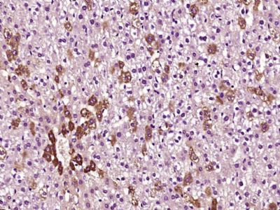
[IF=13.116] Shalaka Wahane. et al. Diversified transcriptional responses of myeloid and glial cells in spinal cord injury shaped by HDAC3 activity. Sci Adv. 2021 Feb;7(9):eabd8811 IHC ; Mouse.
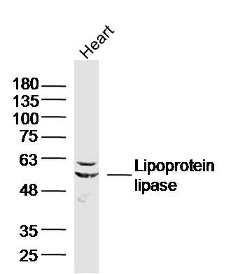
[IF=3.616] Tan W et al. Pyrazinamide alleviates rifampin-induced steatohepatitis in mice by regulating the activities of cholesterol-activated 7α-hydroxylase and lipoprotein lipase. Eur J Pharm Sci
. 2020 Aug 1;151:105402. WB ; Mouse.
[IF=3.514] Li B et al. Resistin up-regulates LPL expression through the PPARγ-dependent PI3K/AKT signaling pathway impacting lipid accumulation in RAW264. 7 macrophages.Cytokine. 2019 Jul;119:168-174. WB ; Mouse.
[IF=2.323] Xiang Yu. et al. Isolation and Identification of Bovine Preadipocytes and Screening of MicroRNAs Associated with Adipogenesis. Animals-Basel. 2020 May;10(5):818 IF ; Bovine.