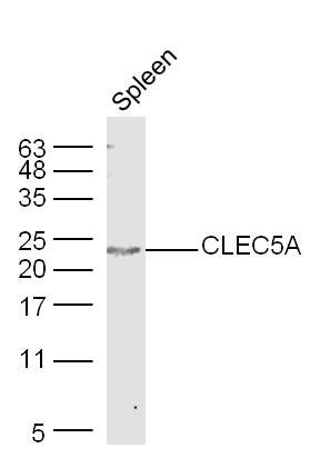
Sample: Spleen (Mouse) Lysate at 40 ug
Primary: Anti-CLEC5A(SL2663R) at 1/300 dilution
Secondary: IRDye800CW Goat Anti-Rabbit IgG at 1/20000 dilution
Predicted band size: 22 kD
Observed band size: 22 kD
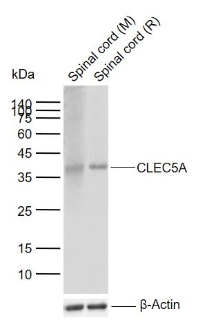
Sample:
Lane 1: Mouse Spinal cord tissue lysates
Lane 2: Rat Spinal cord tissue lysates
Primary: Anti-CLEC5A (SL2663R) at 1/1000 dilution
Secondary: IRDye800CW Goat Anti-Rabbit IgG at 1/20000 dilution
Predicted band size: 22 kDa
Observed band size: 37 kDa
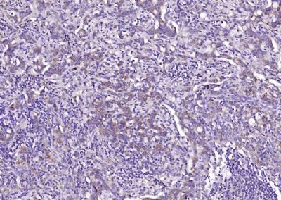
Paraformaldehyde-fixed, paraffin embedded (human gastric carcinoma); Antigen retrieval by boiling in sodium citrate buffer (pH6.0) for 15min; Block endogenous peroxidase by 3% hydrogen peroxide for 20 minutes; Blocking buffer (normal goat serum) at 37°C for 30min; Antibody incubation with (CLEC5A) Polyclonal Antibody, Unconjugated (SL2663R) at 1:200 overnight at 4°C, followed by operating according to SP Kit(Rabbit) (sp-0023) instructionsand DAB staining.
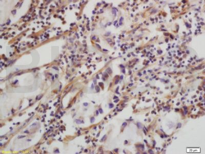
Tissue/cell: human colon carcinoma; 4% Paraformaldehyde-fixed and paraffin-embedded;
Antigen retrieval: citrate buffer ( 0.01M, pH 6.0 ), Boiling bathing for 15min; Block endogenous peroxidase by 3% Hydrogen peroxide for 30min; Blocking buffer (normal goat serum,SLC0005) at 37℃ for 20 min;
Incubation: Anti-CLEC5A/MDL1 Polyclonal Antibody, Unconjugated(SL2663R) 1:200, overnight at 4°C, followed by conjugation to the secondary antibody(SP-0023) and DAB(SLC0010) staining
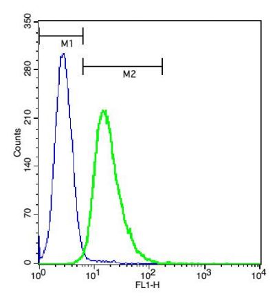
Cell: U937.
Concentration: 1:100.
Incubation: 40 minutes, room temperature.
Host/Blank: U937 cells.
Flow cytometric analysis of Rabbit Anti-CLEC5A antibody (SL2663R) (green) compared with control in the absence of primary antibody (blue) followed by U937 cells.
secondary antibody: Goat Anti-rabbit IgG/FITC antibody (SL0295G-FITC)
|