Home > Product > Antibody > Rabbit Anti-Kir6.2 antibody
ATP sensitive inward rectifier potassium channel 11; Beta cell inward rectifier subunit; mBIR; BIR; HHF 2; HHF2; IKATP; Inward rectifier K(+) channel Inwardly rectifying potassium channel KIR6.2; IRK 11; IRK11; KCNJ11; Kir 6.2; MGC133230; PHHI; Potassium
Cat:
SL2436R
Species Reactivity:
Human,Mouse,(predicted: Rat,Dog,Cow,Rabbit,)
Immunogen:
KLH conjugated synthetic peptide derived from human Kir62:301-390/390
Format:
Liquid
Storage instructions:
Shipped at 4℃. Store at -20 °C for one year. Avoid repeated freeze/thaw cycles.
Concentration:
1mg/ml
Clonality:
Polyclonal
Isotype:
IgG
Applications:
WB=1:500-2000ELISA=1:5000-10000Flow-Cyt=3ug/testIF=1:100-500not yet tested in other applications.optimal dilutions/concentrations should be determined by the end user.
Host:
Rabbit
Product Overview:
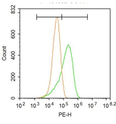
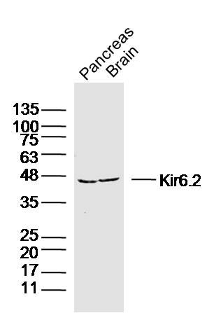
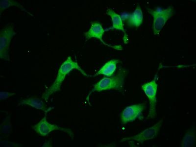
Sample: Pancreas (Mouse) Lysate at 40 ugBrain (Mouse) Lysate at 40 ugPrimary: Anti-Kir6.2 (SL2436R) at 1/300 dilutionSecondary: IRDye800CW Goat Anti-Rabbit IgG at 1/20000 dilutionPredicted band size: 43 kDObserved band size: 45 kDTissue/cell: HeLa cell; 4% Paraformaldehyde-fixed; Triton X-100 at room temperature for 20 min; Blocking buffer (normal goat serum, SLC0005) at 37°C for 20 min; Antibody incubation with (Kir6.2) Polyclonal Antibody, Unconjugated (SL2436R) 1:100, 90 minutes at 37°C; followed by a conjugated Goat Anti-Rabbit IgG antibody (SL0295G-FITC) at 37°C for 90 minutes, DAPI (5ug/ml, blue, SLC0033) was used to stain the cell nuclei.Blank control: A549. Primary Antibody (green line): Rabbit Anti-Kir6.2 antibody (SL2436) Dilution: 3μg /10^6 cells; Isotype Control Antibody (orange line): Rabbit IgG . Secondary Antibody : Goat anti-rabbit IgG-PEDilution: 1μg /test. ProtocolThe cells were incubated in 5%BSA to block non-specific protein-protein interactions for 30 min at at room temperature .Cells stained with Primary Antibody for 30 min at room temperature. The secondary antibody used for 40 min at room temperature. Acquisition of 20,000 events was performed.
More
Unit:
Price: $
Product PDFs
Datasheet:


Potassium channels are present in most mammalian cells, where they participate in a wide range of physiologic responses. The protein encoded by this gene is an integral membrane protein and inward-rectifier type potassium channel. The encoded protein, which has a greater tendency to allow potassium to flow into a cell rather than out of a cell, is controlled by G-proteins and is found associated with the sulfonylurea receptor SUR. Mutations in this gene are a cause of familial persistent hyperinsulinemic hypoglycemia of infancy (PHHI), an autosomal recessive disorder characterized by unregulated insulin secretion. Defects in this gene may also contribute to autosomal dominant non-insulin-dependent diabetes mellitus type II (NIDDM), transient neonatal diabetes mellitus type 3 (TNDM3), and permanent neonatal diabetes mellitus (PNDM). Multiple alternatively spliced transcript variants that encode different protein isoforms have been described for this gene. [provided by RefSeq]

Function:
This receptor is controlled by G proteins. Inward rectifier potassium channels are characterized by a greater tendency to allow potassium to flow into the cell rather than out of it. Their voltage dependence is regulated by the concentration of extracellular potassium; as external potassium is raised, the voltage range of the channel opening shifts to more positive voltages. The inward rectification is mainly due to the blockage of outward current by internal magnesium. Can be blocked by extracellular barium (By similarity). Subunit of ATP-sensitive potassium channels (KATP). Can form cardiac and smooth muscle-type KATP channels with ABCC9. KCNJ11 forms the channel pore while ABCC9 is required for activation and regulation.

Subunit:
Interacts with ABCC8/SUR. Interacts with ABCC9/SUR2.

Subcellular Location:
Cell Membrane; Multi-pass membrane protein

Post-translational modifications:
Phosphorylation by MAPK1 results in changes in channel gating that destabilize the closed states and reduce the ATP sensitivity.

DISEASE:
Familial hyperinsulinemic hypoglycemia 2 (HHF2) [MIM:601820]: Most common cause of persistent hypoglycemia in infancy. Unless early and aggressive intervention is undertaken, brain damage from recurrent episodes of hypoglycemia may occur. Note=The disease is caused by mutations affecting the gene represented in this entry.
Diabetes mellitus, permanent neonatal (PNDM) [MIM:606176]: A rare form of diabetes distinct from childhood-onset autoimmune diabetes mellitus type 1. It is characterized by insulin-requiring hyperglycemia that is diagnosed within the first months of life. Permanent neonatal diabetes requires lifelong therapy. Note=The disease is caused by mutations affecting the gene represented in this entry.
Transient neonatal diabetes mellitus 3 (TNDM3) [MIM:610582]: Neonatal diabetes mellitus, defined as insulin-requiring hyperglycemia within the first month of life, is a rare entity. In about half of the neonates, diabetes is transient and resolves at a median age of 3 months, whereas the rest have a permanent form of diabetes. In a significant number of patients with transient neonatal diabetes mellitus, diabetes type 2 appears later in life. The onset and severity of TNDM3 is variable with childhood-onset diabetes, gestational diabetes or adult-onset diabetes described. Note=The disease is caused by mutations affecting the gene represented in this entry. [DISEASE] Note=Defects in KCNJ11 may contribute to non-insulin-dependent diabetes mellitus (NIDDM), also known as diabetes mellitus type 2.

Similarity:
Belongs to the inward rectifier-type potassium channel (TC 1.A.2.1) family. KCNJ11 subfamily.

SWISS:
Q14654

Gene ID:
3767

Database links:

Entrez Gene: 3767 Human

Entrez Gene: 16514 Mouse

Omim: 600937 Human

SwissProt: Q14654 Human

SwissProt: Q61743 Mouse

Unigene: 248141 Human

Unigene: 333863 Mouse


Picture

Sample:
Pancreas (Mouse) Lysate at 40 ug
Brain (Mouse) Lysate at 40 ug
Primary: Anti-Kir6.2 (SL2436R) at 1/300 dilution
Secondary: IRDye800CW Goat Anti-Rabbit IgG at 1/20000 dilution
Predicted band size: 43 kD
Observed band size: 45 kD
Tissue/cell: HeLa cell; 4% Paraformaldehyde-fixed; Triton X-100 at room temperature for 20 min; Blocking buffer (normal goat serum, SLC0005) at 37°C for 20 min; Antibody incubation with (Kir6.2) Polyclonal Antibody, Unconjugated (SL2436R) 1:100, 90 minutes at 37°C; followed by a conjugated Goat Anti-Rabbit IgG antibody (SL0295G-FITC) at 37°C for 90 minutes, DAPI (5ug/ml, blue, SLC0033) was used to stain the cell nuclei.
Blank control: A549.
Primary Antibody (green line): Rabbit Anti-Kir6.2 antibody (SL2436)
Dilution: 3μg /10^6 cells;
Isotype Control Antibody (orange line): Rabbit IgG .
Secondary Antibody : Goat anti-rabbit IgG-PE
Dilution: 1μg /test.
Protocol
The cells were incubated in 5%BSA to block non-specific protein-protein interactions for 30 min at at room temperature .Cells stained with Primary Antibody for 30 min at room temperature. The secondary antibody used for 40 min at room temperature. Acquisition of 20,000 events was performed.
Product Feedback Wall
Message :
Your Email :
Copyright © 2007-2018 Sunlong Medical All Rights Reserved.