Home > Product > Antibody > Rabbit Anti-AQP7 antibody
aquaporin Protein-7; AQP7; AQP7L; AQP9; AQPap; Aqpap7; Aquaporin 7;Aquaporin-7 like; MGC149555; MGC149556; AQP7_HUMAN.
Cat:
SL2506R
Species Reactivity:
Human,Mouse,(predicted: Rat,Chicken,Dog,Pig,Cow,)
Immunogen:
KLH conjugated synthetic peptide derived from human AQP7:251-342/342<Cytoplasmic>
Format:
Liquid
Storage instructions:
Shipped at 4℃. Store at -20 °C for one year. Avoid repeated freeze/thaw cycles.
Concentration:
1mg/ml
Clonality:
Polyclonal
Isotype:
IgG
Applications:
WB=1:500-2000ELISA=1:5000-10000IHC-P=1:100-500IHC-F=1:100-500Flow-Cyt=2ug/testIF=1:100-500(Paraffin sections need to do antigen repair)not yet tested in other applications.optimal dilutions/concentrations should be determined by the end user.
Host:
Rabbit
Product Overview:
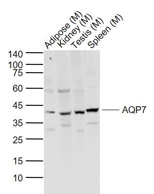
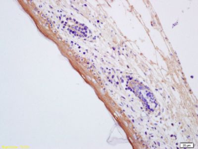
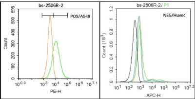
Sample: Lane 1: Adipose (Mouse) Lysate at 40 ugLane 2: Kidney (Mouse) Lysate at 40 ugLane 3: Testis (Mouse) Lysate at 40 ugLane 4: Spleen (Mouse) Lysate at 40 ugPrimary: Anti-AQP7 (SL2506R) at 1/1000 dilutionSecondary: IRDye800CW Goat Anti-Rabbit IgG at 1/20000 dilutionPredicted band size: 37/18 kDObserved band size: 40 kDTissue/cell:Mouse embryos; 4% Paraformaldehyde-fixed and paraffin-embedded; Antigen retrieval: citrate buffer ( 0.01M, pH 6.0 ), Boiling bathing for 15min; Block endogenous peroxidase by 3% Hydrogen peroxide for 30min; Blocking buffer (normal goat serum,SLC0005) at 37℃ for 20 min; Incubation: Anti-AQP7 Polyclonal Antibody, Unconjugated(SL2506R) 1:200, overnight at 4°C, followed by conjugation to the secondary antibody(SP-0023) and DAB(SLC0010) stainingBlack line : Positive blank control (A549); Negative blank control (HUVEC) Green line : Primary Antibody (Rabbit Anti-AQP7 antibody (SL2506R) ) Orange line:Isotype Control Antibody (Rabbit IgG) . Blue line : Secondary Antibody (Goat anti-rabbit IgG-AF488) A549(Positive)and HUVEC(Negative control)cells (black) were incubated in 5% BSA blocking buffer for 30 min at room temperature. Cells were then stained with AQP7 Antibody(SL2506R)at 1:50 dilution in blocking buffer and incubated for 30 min at room temperature, washed twice with 2% BSA in PBS, followed by secondary antibody(blue) incubation for 40 min at room temperature. Acquisitions of 20,000 events were performed. Cells stained with primary antibody (green), and isotype control (orange).Blank control: A549. Primary Antibody (green line): Rabbit Anti-AQP7 antibody (SL2506R) Dilution: 3μg /10^6 cells; Isotype Control Antibody (orange line): Rabbit IgG . Secondary Antibody : Goat anti-rabbit IgG-PEDilution: 3μg /test. ProtocolThe cells were incubated in 5%BSA to block non-specific protein-protein interactions for 30 min at at room temperature .Cells stained with Primary Antibody for 30 min at room temperature. The secondary antibody used for 40 min at room temperature. Acquisition of 20,000 events was performed.
More
Unit:
Price: $
Product PDFs
Datasheet:


Water is a critical component of all living cells. Interestingly, tissue membranes show a great degree of water permeability. Mammalian red cells, renal proximal tubules, and descending thin limb of Henle are extraordinarily permeable to water. Water crosses hydrophobic plasma membranes either by simple diffusion or through a facilitative transport mechanism mediated by special protein "aquaporin". Over the last decade, genes for several members of aquaporin family have been cloned, expressed, and their distribution studied in many tissues. AQP0 or MIP26 (major intrinsic protein 26kD), and Aquaporin 1 (AQP1, purified from red cells) also called CHIP28 (channel forming integral protein, 28kD; 268aa; gene locus 7p14) has been the foundation of the growing family of aquaporin. The lens specific AQP0 represents up to 80% of total lens membrane protein. Defects in MIP26 are cause of autosomal dominant cataract. The cataract Fraser mutation (CATFR or Shriveled) is a transposon induced splicing error that substitutes a long terminal repeat sequence for the C terminus of MIP. The lens opacity mutation (LOP) is an amino acid substitution that inhibits targeting of MIP to the cell membrane.

Function:
Forms a channel for water and glycerol (By similarity).

Subcellular Location:
Membrane; Multi-pass membrane protein.

Similarity:
Belongs to the MIP/aquaporin (TC 1.A.8) family.

SWISS:
O14520

Gene ID:
364

Database links:

Entrez Gene: 364 Human

Entrez Gene: 11832 Mouse

Entrez Gene: 29171 Rat

Omim: 602974 Human

SwissProt: O14520 Human

SwissProt: O54794 Mouse

SwissProt: P51283 Rat

Unigene: 455323 Human

Unigene: 8728 Mouse

Unigene: 11111 Rat


Picture

Sample:
Lane 1: Adipose (Mouse) Lysate at 40 ug
Lane 2: Kidney (Mouse) Lysate at 40 ug
Lane 3: Testis (Mouse) Lysate at 40 ug
Lane 4: Spleen (Mouse) Lysate at 40 ug
Primary: Anti-AQP7 (SL2506R) at 1/1000 dilution
Secondary: IRDye800CW Goat Anti-Rabbit IgG at 1/20000 dilution
Predicted band size: 37/18 kD
Observed band size: 40 kD
Tissue/cell:Mouse embryos; 4% Paraformaldehyde-fixed and paraffin-embedded;
Antigen retrieval: citrate buffer ( 0.01M, pH 6.0 ), Boiling bathing for 15min; Block endogenous peroxidase by 3% Hydrogen peroxide for 30min; Blocking buffer (normal goat serum,SLC0005) at 37℃ for 20 min;
Incubation: Anti-AQP7 Polyclonal Antibody, Unconjugated(SL2506R) 1:200, overnight at 4°C, followed by conjugation to the secondary antibody(SP-0023) and DAB(SLC0010) staining
Black line : Positive blank control (A549); Negative blank control (HUVEC)
Green line : Primary Antibody (Rabbit Anti-AQP7 antibody (SL2506R) )
Orange line:Isotype Control Antibody (Rabbit IgG) .
Blue line : Secondary Antibody (Goat anti-rabbit IgG-AF488)
A549(Positive)and HUVEC(Negative control)cells (black) were incubated in 5% BSA blocking buffer for 30 min at room temperature. Cells were then stained with AQP7 Antibody(SL2506R)at 1:50 dilution in blocking buffer and incubated for 30 min at room temperature, washed twice with 2% BSA in PBS, followed by secondary antibody(blue) incubation for 40 min at room temperature. Acquisitions of 20,000 events were performed. Cells stained with primary antibody (green), and isotype control (orange).
Blank control: A549.
Primary Antibody (green line): Rabbit Anti-AQP7 antibody (SL2506R)
Dilution: 3μg /10^6 cells;
Isotype Control Antibody (orange line): Rabbit IgG .
Secondary Antibody : Goat anti-rabbit IgG-PE
Dilution: 3μg /test.
Protocol
The cells were incubated in 5%BSA to block non-specific protein-protein interactions for 30 min at at room temperature .Cells stained with Primary Antibody for 30 min at room temperature. The secondary antibody used for 40 min at room temperature. Acquisition of 20,000 events was performed.
Product Feedback Wall
Message :
Your Email :
Copyright © 2007-2018 Sunlong Medical All Rights Reserved.