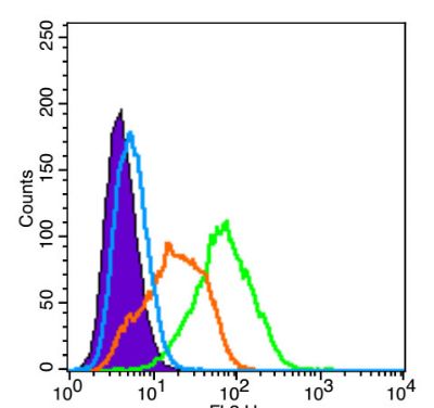
[IF=5.58] Franken, Lars, et al. "Splenic red pulp macrophages are intrinsically superparamagnetic and contaminate magnetic cell isolates." Scientific Reports5 (2015). Mouse.
[IF=2.829] Chen, Xionglin, et al. "Peptide-modified chitosan hydrogels promote skin wound healing by enhancing wound angiogenesis and inhibiting inflammation." Am J Transl Res 9.5 (2017): 2352-2362. IHSLCP ; Mouse.
[IF=2.433] Zhou S et al. High Expression of Angiopoietin-like Protein 4 in Advanced Colorectal Cancer and its Association with Regulatory T Cells and M2 Macrophages. Pathol Oncol Res. 2019 Jul 1. IHSLCP ; Human.
[IF=8.109] Thiago DeSouza-Vieira. et al. Heme Oxygenase-1 Induction by Blood-Feeding Arthropods Controls Skin Inflammation and Promotes Disease Tolerance. Cell Rep. 2020 Oct;33:108317 FC ; Mouse.
[IF=8.897] Liu, Houli. et al. Immunomodulatory hybrid bio-nanovesicle for self-promoted photodynamic therapy. Nano Res. 2022 Feb;:1-10 WB ; Mouse.
[IF=20.722] Meng Lin. et al. CRISPR-based in situ engineering tumor cells to reprogram macrophages for effective cancer immunotherapy. Nano Today. 2022 Feb;42:101359 IF ; Mouse.
[IF=6.922] Shanping Wang. et al. Isosteviol Sodium Exerts Anti-Colitic Effects on BALB/c Mice with Dextran Sodium Sulfate-Induced Colitis Through Metabolic Reprogramming and Immune Response Modulation. J Inflamm Res. 2021; 14: 7107–7130 IF ; Mouse.
[IF=5.501] Haneen S. Dwaib. et al. Phosphorus Supplementation Mitigates Perivascular Adipose Inflammation–Induced Cardiovascular Consequences in Early Metabolic Impairment. J Am Heart Assoc. 2021;0:e023227 FC ; Rat.
[IF=6.244] Yang Chenbo. et al. Relationship Between PTEN and Angiogenesis of Esophageal Squamous Cell Carcinoma and the Underlying Mechanism. Front Oncol. 2021 Nov;0:4638 ICC ; Human.
[IF=3.156] Miaomiao Sun. et al. Tumor-associated macrophages modulate angiogenesis and tumor growth in a xenograft mouse model of multiple myeloma. Leukemia Res. 2021 Nov;110:106709 IHC ; mouse.
[IF=6.353] Luo Haiyun. et al. Concentrated growth factor regulates the macrophage-mediated immune response. Regen Biomater. 2021 Sep;8(6): IF ; mouse.
[IF=5.984] Nina P. Connolly. et al. Elevated fibroblast growth factor-inducible 14 expression transforms proneural-like gliomas into more aggressive and lethal brain cancer. 2021 May 15 IF ; Rat.
[IF=4.171] Rena Fujishima. et al. Similar distribution of orally administered eicosapentaenoic acid and M2 macrophage marker in the hypoperfusion-induced abdominal aortic aneurysm wall. 2021 Mar 29 IHC ; Rat.
[IF=10.317] Yang G et al. A multifunctional anti-inflammatory drug that can specifically target activated macrophages, massively deplete intracellular H2O2, and produce large amounts CO for a highly efficient treatment of osteoarthritis. Biomaterials. 2020 Oct;255:120155. ICF ; Mouse.
[IF=10.317] Yang G et al. A multifunctional anti-inflammatory drug that can specifically target activated macrophages, massively deplete intracellular H 2 O 2, and produce large amounts CO for a highly efficient treatment of osteoarthritis. Biomaterials
. 2020 Oct;255:120155. ICF ; Mouse.
[IF=4.978] Kim MS et al. Enhancement of antitumor effect of radiotherapy via combination with Au@ SiO2 nanoparticles targeted to tumor-associated macrophages. Journal of Industrial and Engineering Chemistry. 2020. Other ; Mouse.
[IF=2.895] Shao N et al. A Multi‐Functional Silicon Nanoparticle Designed for Enhanced Osteoblast Calcification and Related Combination Therapy. Macromol Biosci. 2019 Nov 10:e1900255. FCM&ICF ; Mouse.
[IF=3.616] Moser PT et al. Creation of Laryngeal Grafts from Primary Human Cells and Decellularized Laryngeal Scaffolds. Tissue Eng Part A. 2019 Oct 30. IHSLCP ; beagle.
[IF=2.238] Liu GZ et al.
Aldosterone stimulation mediates cardiac metabolism remodeling via Sirt1/AMPK signaling in canine model.Naunyn Schmiedebergs Arch Pharmacol. 2019 Mar 9. IHSLCP ; Dog.
[IF=5.554] Okonogi N et al. Combination therapy of intravenously injected microglia and radiotherapy prolongs survival in a rat model of spontaneous malignant glioma.International Journal of Radiation Oncology*Biology*Physics. 2018. IHSLCP ; Rat.
[IF=5.97] Rashad S et al. Intracellular S1P Levels Dictate Fate of Different Regions of the
4 Hippocampus following Transient Global Cerebral Ischemia.Neuroscience, 384, 188–202. IHF ; Rat.
[IF=5.29] Protti et al. Bone marrow transplantation modulates tissue macrophage phenotype and enhances cardiac recovery after subsequent acute myocardial infarction. (2016) J.Mol.Cell.Cardiol. 90:120-8 IHC ; Mouse.
[IF=4.12] Tran et al. Modulation of Macrophage Functional Polarity towards Anti-Inflammatory Phenotype with Plasmid DNA Delivery in CD44 Targeting Hyaluronic Acid Nanoparticles. (2015) Sci.Rep. 5:16632 FACS ; Human, Mouse, Rat, Dog, Pig, Horse,.
[IF=4.26] Chen et al. Mesenchymal stem cell-laden anti-inflammatory hydrogel enhances diabetic wound healing. (2015) Sci.Re. 5:18104 IHSLCP ; Mouse.
[IF=5.76] Das, Subhamoy, et al. "Syndesome Therapeutics for Enhancing Diabetic Wound Healing." Advanced Healthcare Materials (2016). IHSLCP ; Mouse.
[IF=0] Dharmakumar, Rohan, and Ivan Cokic. "Assessment of iron deposition post myocardial infarction as a marker of myocardial hemorrhage." U.S. Patent Application No. 14/125,307. IHSLCP ; Dog.
[IF=5.23] Kugo, H. et al. "Adipocyte in vascular wall can induce the rupture of abdominal aortic aneurysm." Sci. Rep. 6, 31268 IHSLCF ; Rat.
[IF=5.04] Drel, Viktor R., et al. "Centrality of bone marrow in the severity of gadolinium-based contrast-induced systemic fibrosis." The FASEB Journal(2016): fj-20300188R. IHSLCP ; Rat.
[IF=7.21] Oh, Jisu, et al. "Deletion of Macrophage Vitamin D Receptor Promotes Insulin Resistance and Monocyte Cholesterol Transport to Accelerate Atherosclerosis in Mice." Cell Reports (2015). Mouse.
[IF=2.46] Wang, Shan, et al. "5-Aminolevulinic Acid–mediated Sonodynamic Therapy Reverses Macrophage and Dendritic Cell Passivity in Murine Melanoma Xenografts." Ultrasound in Medicine & Biology (2014). IHSLCP ; Mouse.
[IF=4.2] Ge, L. P., et al. "Integration of nondegradable polystyrene and degradable gelatin in a core–sheath nanofibrous patch for pelvic reconstruction." International Journal of Nanomedicine 2015:10 3193–641 IHSLCP ; Rat.
[IF=4.26] Riuzzi et al. Levels of S100B protein drive the reparative process in acute muscle injury and muscular dystrophy. (2017) Sci.Re. 7:12537 FCM(FACS) ; Mouse.
[IF=2.745] Gill N et al. The immunophenotype of decidual macrophages in acute atherosis. Am J Reprod Immunol. 2019 Feb 8:e13098. IHF ; Human.
[IF=1.935] Akazawa Y et al. M-CSF Receptor Antagonists Inhibit the Initiation and Progression of Hepatocellular Carcinoma in Mice. Anticancer Res. 2019 Sep;39(9):4787-4794. IHF ; Mouse.
[IF=7.561] Pengfei Li. et al. Comparative Proteomic Analysis of Polarized Human THP-1 and Mouse RAW264.7 Macrophages. Front Immunol. 2021; 12: 700009 IF ; Human, Mouse.
[IF=5.268] An-tian Xu. et al. Effects of strontium-incorporated micro/nano rough titanium surfaces on osseointegration via modulating polarization of macrophages. Colloid Surface B. 2021 Nov;207:111992 IHC ; Rat.
[IF=6.543] Wang Shanping. et al. Isosteviol Sodium Ameliorates Dextran Sodium Sulfate-Induced Chronic Colitis through the Regulation of Metabolic Profiling, Macrophage Polarization, and NF-κB Pathway. Oxid Med Cell Longev. 2022;2022:4636618 IF ; Mouse.
[IF=4.379] Yamazaki, Reiji. et al. Macroscopic detection of demyelinated lesions in mouse PNS with neutral red dye. Sci Rep-Uk. 2021 Aug;11(1):1-11 IF ; Mouse.
[IF=3.623] Sarah Al-Maawi. et al. Multinucleated Giant Cells Induced by a Silk Fibroin Construct Express Proinflammatory Agents: An Immunohistological Study. Materials. 2021 Jan;14(14):4038 IHC ; Rat.
[IF=2.942] Guo Fan. et al. The correlation between tumor-associated macrophage infiltration and progression in cervical carcinoma. Bioscience Rep. 2021 May;41(5):BSR20203145 IHC ; Human.
[IF=2.886] Fan Guo #et al. Tumor-Associated CD163+ M2 Macrophage Infiltration is Highly Associated with PD-L1 Expression in Cervical Cancer. Cancer Manag Res
. 2020 Jul 15;12:5831-5843. IHC ; Human.
[IF=3.496] Philipp T Moser et al. Creation of Laryngeal Grafts from Primary Human Cells and Decellularized Laryngeal Scaffolds. Tissue Eng Part A. 2020 May;26(9-10):543-555. IF ; Rat.
[IF=2.738] Liang Wang et al. Clinical Significance of M1/M2 Macrophages and Related Cytokines in Patients with Spinal Tuberculosis. Dis Markers. 2020 May 20;2020:2509454. IHC ; Human.
[IF=3.03] Xu Y et al. A porcine alveolar macrophage cell line stably expressing CD163 demonstrates virus replication and cytokine secretion characteristics similar to primary alveolar macrophages following PRRSV infection. Vet Microbiol
. 2020 May;244:108690. WB ; pig.
[IF=8.579] Tong L et al. ACT001 reduces the expression of PD-L1 by inhibiting the phosphorylation of STAT3 in glioblastoma. Theranostics. 2020 May 1;10(13):5943-5956. IHC ; Mouse.
[IF=3.382] Luo Q et al. Interleukin-33 Protects Ischemic Brain Injury by Regulating Specific Microglial Activities.Neuroscience. 2018 Aug 10;385:75-89. WB ; Mouse.
[IF=2.088] Wang G et al. PPAR-γ Promotes Hematoma Clearance through Haptoglobin-Hemoglobin-CD163 in a Rat Model of Intracerebral Hemorrhage. Behavioural Neurology, 2018, 1–7. WB ; Rat.
[IF=4.318] Thomas AJ et al. Macrophage CD163 expression in cerebrospinal fluid: association with subarachnoid hemorrhage outcome.J Neurosurg. 2018 Jul 1:1-7. ICF ; Human.
[IF=5.51] Kobori et al. Interleukin-18 Amplifies Macrophage Polarization and Morphological Alteration, Leading to Excessive Angiogenesis. (2018) Front.Immunol. 9:334 FC ; Mouse.
[IF=2.81] Fernandez-Bustamante et al. Brief Glutamine Pretreatment Increases Alveolar Macrophage CD163/Heme Oxygenase-1/p38-MAPK Dephosphorylation Pathway and Decreases Capillary Damage but Not Neutrophil Recruitment in IL-1/LPS-Insufflated Rats. (2015) PLoS.On. 10:e0130764 FCM ; Rat.
[IF=2.476] Yin, Li, Cuifang Chang, and Cunshuan Xu. "Expressions Profiles of the Proteins Associated with Carbohydrate Metabolism in Rat Liver Regeneration." BioMed Research International 2017 (2017). WB ; Rat.
[IF=5] Gerzanich, Volodymyr, et al. "Salutary effects of glibenclamide during the chronic phase of murine experimental autoimmune encephalomyelitis." Journal of Neuroinflammation 14.1 (2017): 177. IHSLCP ; Mouse.
[IF=5.12] Bobi, Joaquim, et al. "Intracoronary Administration of Allogeneic Adipose Tissue–Derived Mesenchymal Stem Cells Improves Myocardial Perfusion But Not Left Ventricle Function, in a Translational Model of Acute Myocardial Infarction." Journal of the American Heart Association 6.5 (2017): e005771. IHSLCP ; Pig.
[IF=3.73] Zeng, Wei-qun, et al. "A New Method to Isolate and Culture Rat Kupffer Cells." PLOS ONE 8.8 (2013): e70832. Rat.
[IF=8.2] Lee, Jin, et al. "Preventive Inhibition of Liver Tumorigenesis by Systemic Activation of Innate Immune Functions." Cell Reports 21.7 (2017): 1870-1882. IHSLCF ; Mouse.
[IF=4.38] Wang, Lei, et al. "A novel nano-copper-bearing stainless steel with reduced Cu2+ release only inducing transient foreign body reaction via affecting the activity of NF-κB and Caspase 3." International Journal of Nanomedicine 10 (2015): 6725. IHSLCP ; Mouse.
