This gene encodes a protein that is necessary for the repair of ultraviolet light-damaged DNA. This protein is the smaller subunit of a heterodimeric protein complex that participates in nucleotide excision repair, and this complex mediates the ubiquitylation of histones H3 and H4, which facilitates the cellular response to DNA damage. This subunit appears to be required for DNA binding. Mutations in this gene cause xeroderma pigmentosum complementation group E, a recessive disease that is characterized by an increased sensitivity to UV light and a high predisposition for skin cancer development, in some cases accompanied by neurological abnormalities. [provided by RefSeq].
Function:
Required for DNA repair. Binds to DDB1 to form the USLVdamaged DNA-binding protein complex (the USLVDDB complex). The USLVDDB complex may recognize USLVinduced DNA damage and recruit proteins of the nucleotide excision repair pathway (the NER pathway) to initiate DNA repair. The USLVDDB complex preferentially binds to cyclobutane pyrimidine dimers (CPD), 6-4 photoproducts (6-4 PP), apurinic sites and short mismatches. Also appears to function as the substrate recognition module for the DCX (DDB1-CUL4-X-box) E3 ubiquitin-protein ligase complex DDB1-CUL4-ROC1 (also known as CUL4-DDB-ROC1 and CUL4-DDB-RBX1). The DDB1-CUL4-ROC1 complex may ubiquitinate histone H2A, histone H3 and histone H4 at sites of USLVinduced DNA damage. The ubiquitination of histones may facilitate their removal from the nucleosome and promote subsequent DNA repair. The DDB1-CUL4-ROC1 complex also ubiquitinates XPC, which may enhance DNA-binding by XPC and promote NER. Isoform D1 and isoform D2 inhibit USLVdamaged DNA repair.
Subunit:
Component of the USLVDDB complex which includes DDB1 and DDB2. The USLVDDB complex interacts with monoubiquitinated histone H2A and binds to XPC via the DDB2 subunit. Component of the DCX (DDB1-CUL4-X-box) E3 ubiquitin-protein ligase complex DDB1-CUL4-ROC1 (also known as CUL4-DDB-ROC1 and CUL4-DDB-RBX1), which includes CUL4A or CUL4B, DDB1, DDB2 and RBX1. DDB2 may function as the substrate recognition module within this complex. The DDB1-CUL4-ROC1 complex may associate with the COP9 signalosome, and this inhibits the E3 ubiquitin-protein ligase activity of the complex. A large number of other DCX complexes may also exist in which an alternate substrate targeting subunit replaces DDB2. These targeting subunits are generally known as DCAF (DDB1- and CUL4-associated factor) or CDW (CUL4-DDB1-associated WD40-repeat) proteins. Isoform D1 and isoform D2 do not interact with DDB1.
Subcellular Location:
Nucleus. Note=Accumulates at sites of DNA damage following UV irradiation.
Tissue Specificity:
Ubiquitously expressed; with highest levels in corneal endothelium and lowest levels in brain. Isoform D1 is highly expressed in brain and heart. Isoform D2, isoform D3 and isoform D4 are weakly expressed.
Post-translational modifications:
Phosphorylation by ABL1 negatively regulate USLVDDB activity (By similarity).
Ubiquitinated by CUL4A in response to UV irradiation. Ubiquitination appears to both impair DNA-binding and promotes ubiquitin-dependent proteolysis. Degradation of DDB2 at sites of DNA damage may be a prerequisite for their recognition by XPC and subsequent repair. CUL4A-mediated degradation appears to be promoted by ABL1.
DISEASE:
Xeroderma pigmentosum complementation group E (XP-E) [MIM:278740]: An autosomal recessive pigmentary skin disorder characterized by solar hypersensitivity of the skin, high predisposition for developing cancers on areas exposed to sunlight and, in some cases, neurological abnormalities. The skin develops marked freckling and other pigmentation abnormalities. XP-E patients show a mild phenotype with minimal or no neurologic features. Note=The disease is caused by mutations affecting the gene represented in this entry.
Similarity:
Belongs to the WD repeat DDB2/WDR76 family.
Contains 7 WD repeats.
SWISS:
Q92466
Gene ID:
1643
Database links:
Entrez Gene: 519357 Cow
Entrez Gene: 1643 Human
Entrez Gene: 107986 Mouse
Omim: 600811 Human
SwissProt: Q0VBY8 Cow
SwissProt: Q92466 Human
SwissProt: Q99J79 Mouse
Unigene: 446564 Human
Unigene: 700338 Human
Unigene: 260747 Mouse
Unigene: 389334 Mouse
| Picture |
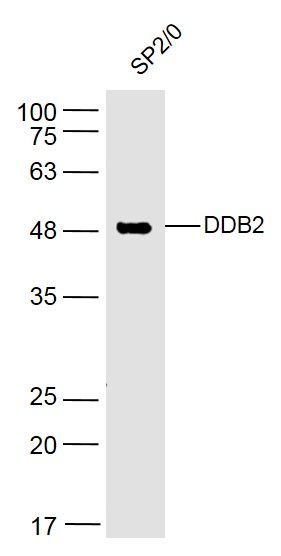
Sample:
SP2/0(Human) Cell Lysate at 30 ug
Primary: Anti- DDB2 (SL2599R) at 1/300 dilution
Secondary: IRDye800CW Goat Anti-Rabbit IgG at 1/20000 dilution
Predicted band size: 48 kD
Observed band size: 48 kD
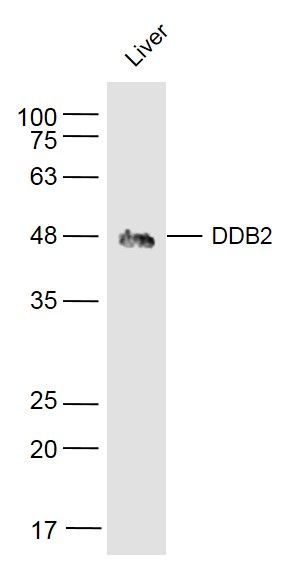
Sample:
Liver (Mouse) Lysate at 40 ug
Primary: Anti- DDB2 (SL2599R) at 1/300 dilution
Secondary: IRDye800CW Goat Anti-Rabbit IgG at 1/20000 dilution
Predicted band size: 48 kD
Observed band size: 48 kD
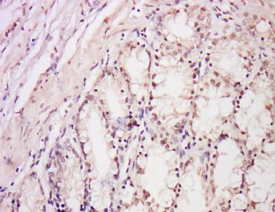
Tissue/cell: Rat intestine tissue; 4% Paraformaldehyde-fixed and paraffin-embedded;
Antigen retrieval: citrate buffer ( 0.01M, pH 6.0 ), Boiling bathing for 15min; Block endogenous peroxidase by 3% Hydrogen peroxide for 30min; Blocking buffer (normal goat serum,SLC0005) at 37℃ for 20 min;
Incubation: Anti-DDB2 Polyclonal Antibody, Unconjugated(SL2599R) 1:200, overnight at 4°C, followed by conjugation to the secondary antibody(SP-0023) and DAB(SLC0010) staining
|
|
|