Home > Product > Antibody > Rabbit Anti-RUNX1 antibody
Acute myeloid leukemia 1; Acute myeloid leukemia 1 protein; alpha subunit core binding factor; AML 1; AML1 EVI 1; Aml1 oncogene; AMLCR 1; AMLCR1; CBFA 2; CBFA2; Core binding factor alpha 2 subunit; Core binding factor runt domain alpha subunit 2; EVI 1; E
Cat:
SL2744R
Species Reactivity:
Human,Mouse,(predicted: Rat,Dog,Cow,Horse,Rabbit,)
Immunogen:
KLH conjugated synthetic peptide derived from human RUNX1:101-200/453
Format:
Liquid
Storage instructions:
Shipped at 4℃. Store at -20 °C for one year. Avoid repeated freeze/thaw cycles.
Concentration:
1mg/ml
Clonality:
Polyclonal
Isotype:
IgG
Applications:
WB=1:500-2000ELISA=1:5000-10000Flow-Cyt=1ug/Testnot yet tested in other applications.optimal dilutions/concentrations should be determined by the end user.
Host:
Rabbit
Product Overview:
Sample: Thymus (Mouse) Lysate at 40 ugPrimary: Anti-RUNX1 (SL2744R) at 1/300 dilutionSecondary: IRDye800CW Goat Anti-Rabbit IgG at 1/20000 dilutionPredicted band size: 50 kDObserved band size: 50 kDSample: Spleen (Mouse) Lysate at 40 ugPrimary: Anti-RUNX1 (SL2744R) at 1/300 dilutionSecondary: IRDye800CW Goat Anti-Rabbit IgG at 1/20000 dilutionPredicted band size: 50 kDObserved band size: 50 kDBlank control(black line):Molt4. Primary Antibody (green line): Rabbit Anti-RUNX1 antibody (SL2744R) Dilution:1ug/Test; Secondary Antibody(white blue line): Goat anti-rabbit IgG-AF488Dilution: 0.5ug/Test. Isotype control(orange line): Normal Rabbit IgGProtocolThe cells were fixed with 4% PFA (10min at room temperature)and then permeabilized with 90% ice-cold methanol for 20 min at -20℃, The cells were then incubated in 5%BSA to block non-specific protein-protein interactions for 30 min at room temperature .Cells stained with Primary Antibody for 30 min at room temperature. The secondary antibody used for 40 min at room temperature. Acquisition of 20,000 events was performed.Blank control:A431. Primary Antibody (green line): Rabbit Anti-RUNX1 antibody (SL2744R) Dilution: 1μg /10^6 cells; Isotype Control Antibody (orange line): Rabbit IgG . Secondary Antibody : Goat anti-rabbit IgG-AF647Dilution: 1μg /test. ProtocolThe cells were fixed with 4% PFA (10min at room temperature)and then permeabilized with 90% ice-cold methanol for 20 min at-20℃. The cells were then incubated in 5%BSA to block non-specific protein-protein interactions for 30 min at at room temperature .Cells stained with Primary Antibody for 30 min at room temperature. The secondary antibody used for 40 min at room temperature. Acquisition of 20,000 events was performed.Blank control(black line):Molt4. Primary Antibody (green line): Rabbit Anti-RUNX1 antibody (SL2744R) Dilution:1ug/Test; Secondary Antibody(white blue line): Goat anti-rabbit IgG-AF488Dilution: 0.5ug/Test. Isotype control(orange line): Normal Rabbit IgGProtocolThe cells were fixed with 4% PFA (10min at room temperature)and then permeabilized with 90% ice-cold methanol for 20 min at -20℃, The cells were then incubated in 5%BSA to block non-specific protein-protein interactions for 30 min at room temperature .Cells stained with Primary Antibody for 30 min at room temperature. The secondary antibody used for 40 min at room temperature. Acquisition of 20,000 events was performed.
More
Unit:
Price: $
Product PDFs
Datasheet:


AML1/Runx1 binds DNA as a monomer and through the Runt domain. DNA binding is increased by heterodimerization with CSFB. Isoform AML1L can neither bind DNA nor heterodimerize and interferes with the transactivation activity of AML1/Runx1. CSF binds to the core site, 5'-PYGPYGGT-3', of a number of enhancers and promoters, including murine leukemia virus, polyomavirus enhancer, T cell receptor enhancers, LCK, IL3 and GMCSF promoters. The alpha subunit binds DNA and appears to have a role in the development of normal hematopoiesis. AML1/Runx1 is expressed in a wide variety of tissues and is expressed at the highest levels in thymus, bone marrow and peripheral blood. Defects in AML1/Runx1 are the cause of familial platelet disorder with associated myeloid malignancy, an autosomal dominant disease characterized by qualitative and quantitative platelet defects, and propensity to develop acute myelogenous leukemia。

Function:
CSF binds to the core site, 5'-PYGPYGGT-3', of a number of enhancers and promoters, including murine leukemia virus, polyomavirus enhancer, T-cell receptor enhancers, LCK, IL-3 and GM-CSF promoters. The alpha subunit binds DNA and appears to have a role in the development of normal hematopoiesis. Isoform AML-1L interferes with the transactivation activity of RUNX1. Acts synergistically with ELF4 to transactivate the IL-3 promoter and with ELF2 to transactivate the mouse BLK promoter. Inhibits KAT6B-dependent transcriptional activation.

Subunit:
Heterodimer with CSFB. RUNX1 binds DNA as a monomer and through the Runt domain. DNA-binding is increased by heterodimerization. Isoform AML-1L can neither bind DNA nor heterodimerize. Interacts with TLE1 and ALYREF/THOC4. Interacts with ELF1, ELF2 and SPI1. Interacts via its Runt domain with the ELF4 N-terminal region. Interaction with ELF2 isoform 2 (NERF-1a) may act to repress RUNX1-mediated transactivation. Interacts with KAT6A and KAT6B. Interacts with SUV39H1, leading to abrogation of transactivating and DNA-binding properties of RUNX1. Interacts with YAP1. Interacts with HIPK2 (By similarity). Interaction with CDK6 prevents myeloid differentiation, reducing its transcription transactivation activity.

Subcellular Location:
Nucleus.

Tissue Specificity:
Expressed in all tissues examined except brain and heart. Highest levels in thymus, bone marrow and peripheral blood.

Post-translational modifications:
Phosphorylated in its SLCterminus upon IL-6 treatment. Phosphorylation enhances interaction with KAT6A.
Methylated.
Phosphorylated in Ser-249 Thr-273 and Ser-276 by HIPK2 when associated with CSFB and DNA. This phosphorylation promotes subsequent EP300 phosphorylation.

DISEASE:
Note=A chromosomal aberration involving RUNX1/AML1 is a cause of chronic myelogenous leukemia (CML). Translocation t(3;21)(q26;q22) with EAP or MECOM.
Note=A chromosomal aberration involving RUNX1/AML1 is found in childhood acute lymphoblastic leukemia (ALL). Translocation t(12;21)(p13;q22) with TEL. The translocation fuses the 3'-end of TEL to the alternate 5'-exon of AML-1H.
Note=A chromosomal aberration involving RUNX1 is found in acute leukemia. Translocation t(11,21)(q13;q22) that forms a MACROD1-RUNX1 fusion protein.
Defects in RUNX1 are the cause of familial platelet disorder with associated myeloid malignancy (FPDMM) [MIM:601399]. FPDMM is an autosomal dominant disease characterized by qualitative and quantitative platelet defects, and propensity to develop acute myelogenous leukemia.
Note=A chromosomal aberration involving RUNX1/AML1 is found in therapy-related myeloid malignancies. Translocation t(16;21)(q24;q22) that forms a RUNX1-CSFA2T3 fusion protein.
Note=A chromosomal aberration involving RUNX1/AML1 is a cause of chronic myelomonocytic leukemia. Inversion inv(21)(q21;q22) with USP16.

Similarity:
Contains 1 Runt domain.

SWISS:
Q01196

Gene ID:
861

Database links:

Entrez Gene: 861 Human

Entrez Gene: 12394 Mouse

Entrez Gene: 50662 Rat

Omim: 151385 Human

SwissProt: Q01196 Human

SwissProt: Q03347 Mouse

SwissProt: Q63046 Rat

Unigene: 149261 Human

Unigene: 612648 Human

Unigene: 4081 Mouse

Unigene: 470227 Mouse

Unigene: 11201 Rat


Picture

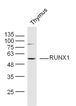
Sample: Thymus (Mouse) Lysate at 40 ug
Primary: Anti-RUNX1 (SL2744R) at 1/300 dilution
Secondary: IRDye800CW Goat Anti-Rabbit IgG at 1/20000 dilution
Predicted band size: 50 kD
Observed band size: 50 kD
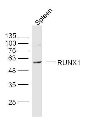
Sample: Spleen (Mouse) Lysate at 40 ug
Primary: Anti-RUNX1 (SL2744R) at 1/300 dilution
Secondary: IRDye800CW Goat Anti-Rabbit IgG at 1/20000 dilution
Predicted band size: 50 kD
Observed band size: 50 kD
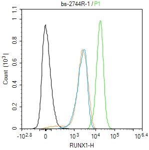
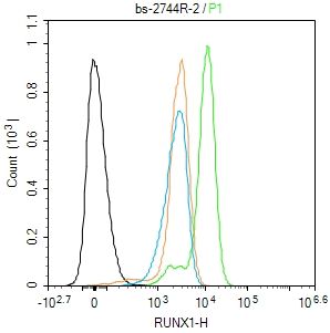
Blank control(black line):Molt4.
Primary Antibody (green line): Rabbit Anti-RUNX1 antibody (SL2744R)
Dilution:1ug/Test;
Secondary Antibody(white blue line): Goat anti-rabbit IgG-AF488
Dilution: 0.5ug/Test.
Isotype control(orange line): Normal Rabbit IgG
Protocol
The cells were fixed with 4% PFA (10min at room temperature)and then permeabilized with 90% ice-cold methanol for 20 min at -20℃, The cells were then incubated in 5%BSA to block non-specific protein-protein interactions for 30 min at room temperature .Cells stained with Primary Antibody for 30 min at room temperature. The secondary antibody used for 40 min at room temperature. Acquisition of 20,000 events was performed.
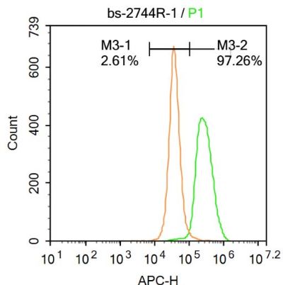
Blank control:A431.
Primary Antibody (green line): Rabbit Anti-RUNX1 antibody (SL2744R)
Dilution: 1μg /10^6 cells;
Isotype Control Antibody (orange line): Rabbit IgG .
Secondary Antibody : Goat anti-rabbit IgG-AF647
Dilution: 1μg /test.
Protocol
The cells were fixed with 4% PFA (10min at room temperature)and then permeabilized with 90% ice-cold methanol for 20 min at-20℃. The cells were then incubated in 5%BSA to block non-specific protein-protein interactions for 30 min at at room temperature .Cells stained with Primary Antibody for 30 min at room temperature. The secondary antibody used for 40 min at room temperature. Acquisition of 20,000 events was performed.
Blank control(black line):Molt4.
Primary Antibody (green line): Rabbit Anti-RUNX1 antibody (SL2744R)
Dilution:1ug/Test;
Secondary Antibody(white blue line): Goat anti-rabbit IgG-AF488
Dilution: 0.5ug/Test.
Isotype control(orange line): Normal Rabbit IgG
Protocol
The cells were fixed with 4% PFA (10min at room temperature)and then permeabilized with 90% ice-cold methanol for 20 min at -20℃, The cells were then incubated in 5%BSA to block non-specific protein-protein interactions for 30 min at room temperature .Cells stained with Primary Antibody for 30 min at room temperature. The secondary antibody used for 40 min at room temperature. Acquisition of 20,000 events was performed.
Product Feedback Wall
Message :
Your Email :
Copyright © 2007-2018 Sunlong Medical All Rights Reserved.