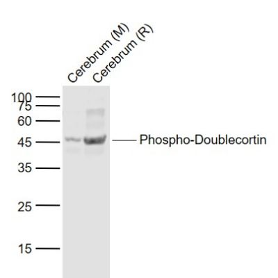
Sample:
Lane 1: Cerebrum (Mouse) Lysate at 40 ug
Lane 2: Cerebrum (Rat) Lysate at 40 ug
Primary: Anti-Phospho-Doublecortin (Ser47) (SL3113R) at 1/1000 dilution
Secondary: IRDye800CW Goat Anti-Rabbit IgG at 1/20000 dilution
Predicted band size: 45 kD
Observed band size: 45 kD
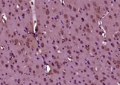
Paraformaldehyde-fixed, paraffin embedded (Rat brain); Antigen retrieval by boiling in sodium citrate buffer (pH6.0) for 15min; Block endogenous peroxidase by 3% hydrogen peroxide for 20 minutes; Blocking buffer (normal goat serum) at 37°C for 30min; Antibody incubation with (Phospho-Doublecortin (Ser128)) Polyclonal Antibody, Unconjugated (SL3113R) at 1:400 overnight at 4°C, followed by operating according to SP Kit(Rabbit) (sp-0023) instructionsand DAB staining.
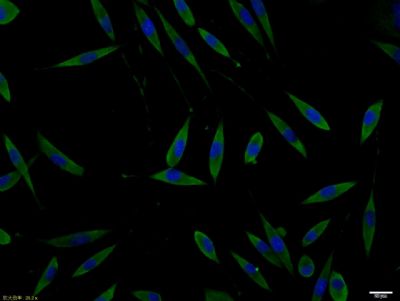
SHSY5Y cell; 4% Paraformaldehyde-fixed; Triton X-100 at room temperature for 20 min; Blocking buffer (normal goat serum, SLC0005) at 37°C for 20 min; Antibody incubation with (Phospho-Doublecortin (Ser128)) polyclonal Antibody, Unconjugated (SL3113R) 1:100, 90 minutes at 37°C; followed by a conjugated Goat Anti-Rabbit IgG antibody at 37°C for 90 minutes, DAPI (blue, C02-04002) was used to stain the cell nuclei.
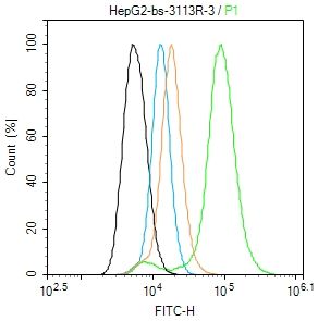
Blank control (black line): HepG2(black) (The cells were fixed with 2% paraformaldehyde (10 min) , then permeabilized with PBST for 30 min on room temperature)
Primary Antibody (green line): Rabbit Anti-Phospho-Doublecortin(Ser128)(SL3113R) ;
Dilution: 1μg /10^6 cells;
Isotype Control Antibody (orange line): Rabbit IgG .
Secondary Antibody (white blue line): Goat anti-rabbit IgG-FITC;Dilution: 1μg /test.
|