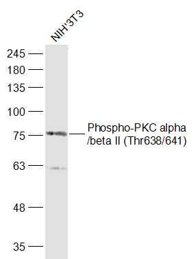
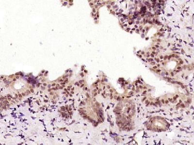
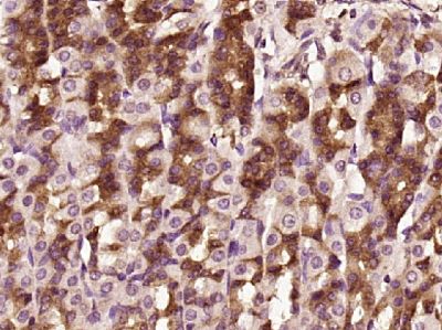
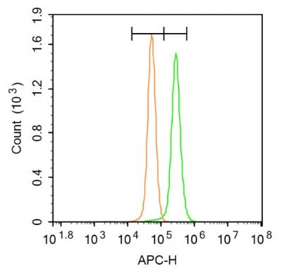
[IF=6.543] Fan Kexia. et al. Drp1-Mediated Mitochondrial Metabolic Dysfunction Inhibits the Tumor Growth of Pituitary Adenomas. Oxid Med Cell Longev. 2022;2022:5652586 WB ; Mouse.
[IF=3.5] Wen-Chien Cheng. et al. Pendulone induces apoptosis via the ROS-mediated ER-stress pathway in human non-small cell lung cancer cells. Toxicol In Vitro. 2022 Jun;81:105346 WB ; Human.
[IF=4.057] Li et al. Apoptosis Induction by Iron Radiation via Inhibition of Autophagy in Trp53+/- Mouse Testes: Is Chronic Restraint-Induced Stress a Modifying Factor?. (2018) Int.J.Biol.Sci. 14:1109-1121 WB ; Mice.
[IF=3.457] Ju Y et al. Protective effects of Astragaloside IV on endoplasmic reticulum stress-induced renal tubular epithelial cells apoptosis in type 2 diabetic nephropathy rats. (2019)Biomedicine & Pharmacotherapy. Jan;109:84-92. WB ; Rat.
[IF=25.606] Raines, Lydia N.. et al. PERK is a critical metabolic hub for immunosuppressive function in macrophages. Nat Immunol. 2022 Feb;23(3):431-445 FC,WB ; Mouse.
[IF=5.118] I-Ta Lu. et al. (−)-Agelasidine A Induces Endoplasmic Reticulum Stress-Dependent Apoptosis in Human Hepatocellular Carcinoma. Mar Drugs. 2022 Feb;20(2):109 WB ; Human.
[IF=3.444] Chang, Lei. et al. Attenuation of Activated eIF2α Signaling by ISRIB Treatment After Spinal Cord Injury Improves Locomotor Function. 2021 Oct 13 WB ; Mice.
[IF=3.159] Tao Tu. et al. Dietary ω-3 fatty acids reduced atrial fibrillation vulnerability via attenuating myocardial endoplasmic reticulum stress and inflammation in a canine model of atrial fibrillation. J Cardiol. 2021 Oct;: WB ; Dog.
[IF=6.268] Nadya Al-Yacoub. et al. Mutation in FBXO32 causes dilated cardiomyopathy through up-regulation of ER-stress mediated apoptosis. Commun Biol. 2021 Jul;4(1):1-12 WB ; Human.
[IF=4.96] Wei Shi. et al. Identification of dihydrotanshinone I as an ERp57 inhibitor with anti-breast cancer properties via the UPR pathway. Biochem Pharmacol. 2021 Aug;190:114637 WB ; Human.
[IF=6.284] Yujie Zhong. et al. Inhibition of ER stress attenuates kidney injury and apoptosis induced by 3-MCPD via regulating mitochondrial fission/fusion and Ca 2+ homeostasis. 2021 Mar 02 WB ; Rat.
[IF=4.872] Yu Wang. et al. Zinc offers splenic protection through suppressing PERK/IRE1-driven apoptosis pathway in common carp (Cyprinus carpio) under arsenic stress. Ecotox Environ Safe. 2021 Jan;208:111473 WB ; Fish.
[IF=5.014] Ji-Eun Kim. et al. Epigallocatechin-3-Gallate and PEDF 335 Peptide, 67LR Activators, Attenuate Vasogenic Edema, and Astroglial Degeneration Following Status Epilepticus. Antioxidants-Basel. 2020 Sep;9(9):854 WB,IHC ; Rat.
[IF=4.096] Xiaowei Sun. et al. Acupuncture protects against cerebral ischemia–reperfusion injury via suppressing endoplasmic reticulum stress-mediated autophagy and apoptosis. Mol Med. 2020 Dec;26(1):1-14 IF ; Rat.
[IF=3.606] Xuejiao Zhou. et al. The novel ALK inhibitor ZX‐29 induces apoptosis through inhibiting ALK and inducing ROS‐mediated endoplasmic reticulum stress in Karpas299 cells. 2020 Nov 02 WB ; Human.
[IF=3.347] Yan Luoet al. Dihydroartemisinin exposure impairs porcine ovarian granulosa cells by activating PERK-eIF2α-ATF4 through endoplasmic reticulum stress. Toxicol Appl Pharmacol
. 2020 Sep 15;403:115159. WB ; pig.
[IF=3.184] Bingwei Yang et al. Polychlorinated Biphenyl Quinone Promotes Atherosclerosis through Lipid Accumulation and Endoplasmic Reticulum Stress via CD36. Chem Res Toxicol. 2020 Jun 15;33(6):1497-1507. WB ; Mouse.
[IF=3.858] Li,et al.Silver nanoparticles induce SH-SY5Y cell apoptosis via endoplasmic reticulum- and mitochondrial pathways that lengthen endoplasmic reticulum-mitochondria contact sites and alter inositol-3-phosphate receptor function.(2018) Toxicology Letters. 285:156-167. WB ; Human.
[IF=4.32] Yan et al. Cytotoxicity of CdTe quantum dots in human umbilical vein endothelial cells: the involvement of cellular uptake and induction of pro-apoptotic endoplasmic reticulum stress. (2016) Int.J.Nanomedicine. 11:529-42 WB ; Human.
[IF=3.7] Mak, Shiu-Kwong, et al. "Tetramethylpyrazine suppresses angiotensin II-induced soluble epoxide hydrolase expression in coronary endothelium via anti-ER stress mechanism." Toxicology and Applied Pharmacology (2017). WB ; Pig.
[IF=9.462] Santanam et al. Atg7 cooperates with Pten loss to drive prostate cancer tumor growth. (2016) Genes.De. 30:399-407 WB ; Mouse.
[IF=3.63] Aslan, Mutay, et al. "Inhibition of Neutral Sphingomyelinase Decreases Elevated Levels of Inducible Nitric Oxide Synthase and Apoptotic Cell Death in Ocular Hypertensive Rats." Toxicology and Applied Pharmacology (2014). WB ; Rat.
[IF=4.196] Kao et al. Opposite Regulation of CHOP and GRP78 and Synergistic Apoptosis Induction by Selenium Yeast and Fish Oil via AMPK Activation in Lung Adenocarcinoma Cells. (2018) Nutrients.ct 8;10(10). WB ; Human.
[IF=4.65] Yu, H., et al. "Gypenoside Protects against Myocardial Ischemia-Reperfusion Injury by Inhibiting Cardiomyocytes Apoptosis via Inhibition of CHOP Pathway and Activation of PI3K/Akt Pathway In Vivo and In Vitro."Cellular Physiology and Biochemistry 39.1 (2016): 123-136. WB ; Rat.
[IF=1.665] Rui Zhang. et al. Apigetrin ameliorates streptozotocin-induced pancreatic β-cell damages via attenuating endoplasmic reticulum stress. In Vitro Cell Dev-An. 2020 Sep;56(8):622-634 WB ; Rat.
[IF=2.614] Bingbing Ma. et al. Dietary taurine supplementation ameliorates muscle loss in chronic heat stressed broilers via suppressing the perk signaling and reversing endoplasmic reticulum‐stress‐induced apoptosis. 2020 Oct 10 IP ; Chicken.
[IF=2.629] Li Yi-Ming. et al. Procyanidin B2 Alleviates Palmitic Acid-Induced Injury in HepG2 Cells via Endoplasmic Reticulum Stress Pathway. Evid-Based Compl Alt. 2021;2021:8920757 WB ; Human.
[IF=5.717] Jianlong Du. et al. FXR, a Key Regulator of Lipid Metabolism, Is Inhibited by ER Stress-Mediated Activation of JNK and p38 MAPK in Large Yellow Croakers (Larimichthys crocea) Fed High Fat Diets. Nutrients. 2021 Dec;13(12):4343 WB ; Fish.
[IF=2.043] Chen Meixue. et al. SMYD1 alleviates septic myocardial injury by inhibiting endoplasmic reticulum stress. Biosci Biotech Bioch. 2021 Oct;: WB ; Rat.
[IF=7.376] Fang Dong. et al. Trimethylamine N-oxide promotes hyperoxaluria-induced calcium oxalate deposition and kidney injury by activating autophagy. Free Radical Bio Med. 2021 Nov;: WB ; Mouse,Human.
[IF=6.081] Ji-Eun Kim. et al. AMPA Receptor Antagonists Facilitate NEDD4-2-Mediated GRIA1 Ubiquitination by Regulating PP2B-ERK1/2-SGK1 Pathway in Chronic Epilepsy Rats. Biomedicines. 2021 Aug;9(8):1069 WB ; Rat.
[IF=41.845] Ashley R. Helseth. et al. Cholinergic neurons constitutively engage the ISR for dopamine modulation and skill learning in mice. Science. 2021 Apr;372(6540): IHC ; Mouse.
[IF=2.092] Zhang Hao. et al. Protective effects of pterostilbene against hepatic damage, redox imbalance, mitochondrial dysfunction, and endoplasmic reticulum stress in weanling piglets. J Anim Sci. 2020 Oct;98(10): WB ; Pig.
[IF=3.038] Shaobin Yang. et al. Neuronostatin Promotion Soluble Aβ1-42 Oligomers: Induced Dysfunctional Brain Glucose Metabolism in Mice. Neurochem Res. 2020 Oct;45(10):2474-2486 WB ; Mouse.
[IF=5.546] Donghui Yang. et al. Melatonin alleviates LPS‐induced endoplasmic reticulum stress and inflammation in spermatogonial stem cells. J Cell Physiol. 2021 May;236(5):3536-3551 WB,IF,IHC ; Mouse.
[IF=5.778] Yuting Wang. et al. Polybrominated diphenyl ethers quinone-induced intracellular protein oxidative damage triggers ubiquitin-proteasome and autophagy-lysosomal system activation in LO2 cells. Chemosphere. 2021 Jul;275:130034 WB ; Human.
[IF=2.971] Wenfeng Gouet al. Ursolic acid derivative UA232 evokes apoptosis of lung cancer cells induced by endoplasmic reticulum stress. Pharm Biol
. 2020 Dec;58(1):707-715. WB ; Human.
[IF=5.656] Kim JE et al. TRPC6-Mediated ERK1/2 Activation Increases Dentate Granule Cell Resistance to Status Epilepticus Via Regulating Lon Protease-1 Expression and Mitochondrial Dynamics. Cells. 2019 Nov 1;8(11). pii: E1376. WB ; Rat.
[IF=4.868] Cui J et al. Acetaldehyde Induces Neurotoxicity In Vitro via Oxidative Stress-and Ca2. Oxid Med Cell Longev. 2019 Jan 9;2019:2593742. WB ; Mouse.
[IF=3.125] Liu.C. et al. Dexmedetomidine alleviates cerebral ischemia-reperfusion injury by inhibiting
endoplasmic reticulum stress dependent apoptosis through the PERK-CHOPCaspase-11
pathway.(2018) Brain Research WB ; Rat.
[IF=2.776] He Q et al. Titanium dioxide nanoparticles induce mouse hippocampal neuron apoptosis via oxidative stress- and calcium imbalance-mediated endoplasmic reticulum stress. Environ Toxicol Pharmacol. 2018 Oct;63:6-15. WB ; Mouse.
[IF=3.226] Terzuoli,et al.Involvement of Bradykinin B2 Receptor in Pathological Vascularization in Oxygen-Induced Retinopathy in Mice and Rabbit Cornea.(2018) International Journal of Molecular Sciences. 19:. WB ; Mouse.
[IF=5.1] Gerbino, Andrea, et al. "Functional Characterization of a Novel Truncating Mutation in Lamin A/C Gene in a Family with a Severe Cardiomyopathy with Conduction Defects." Cellular Physiology and Biochemistry 44.4 (2017): 1559-1577. WB ; Human.
[IF=3.108] Yan, Jiting, et al. "Catalpol prevents alteration of cholesterol homeostasis in non-alcoholic fatty liver disease via attenuating endoplasmic reticulum stress and NOX4 over-expression." RSC Advances 7.2 (2017): 1161-1176. WB ; Human.
[IF=4.94] Carmosino, Monica, et al. "The expression of Lamin A mutant R321X leads to endoplasmic reticulum stress with aberrant Ca2+ handling." Journal of Cellular and Molecular Medicine (2016). WB, IF(ICC) ; Human.
[IF=0] Wang, Yu, et al. "Tanshinone II A Relieves Adriamycin-induced Myocardial Injury in Rat Model." International Journal of Chemistry 8.1 (2016): 40. WB ; Rat.
[IF=4.19] Xu, Demei, et al. "Polychlorinated biphenyl quinone induces endoplasmic reticulum stress, unfolded protein response and calcium release." Chemical Research in Toxicology (2015). WB ; Human.
[IF=5.27] Kucuksayan, Ertan, et al. "Neutral Sphingomyelinase Inhibition Decreases ER Stress-Mediated Apoptosis and Inducible Nitric Oxide Synthase in Retinal Pigment Epithelial Cells." Free Radical Biology and Medicine (2014). WB ; Human.
[IF=2.33] He, Yihuai, et al. "Sustained endoplasmic reticulum stress inhibits hepatocyte proliferation via downregulation of c-Met expression." Molecular and Cellular Biochemistry (2014): 1-8. WB ; Human.


