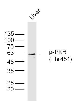
Sample:
Liver (Mouse) Lysate at 40 ug
Primary: Anti- p-PKR (Thr451) (SL3336R) at 1/300 dilution
Secondary: IRDye800CW Goat Anti-Rabbit IgG at 1/20000 dilution
Predicted band size: 62 kD
Observed band size: 62 kD
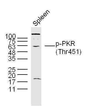
Sample:
Spleen (Mouse) Lysate at 40 ug
Primary: Anti- p-PKR(Thr451) (SL3336R)at 1/300 dilution
Secondary: IRDye800CW Goat Anti-Rabbit IgG at 1/20000 dilution
Predicted band size: 62 kD
Observed band size: 62 kD
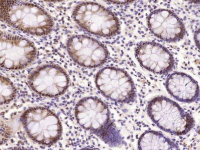
Paraformaldehyde-fixed, paraffin embedded (Human colon carcinoma); Antigen retrieval by microwave in sodium citrate buffer (pH6.0) ; Block endogenous peroxidase by 3% hydrogen peroxide for 30 minutes; Blocking buffer (3% BSA) at RT for 30min; Antibody incubation with (Phospho-PKR (Thr451)) Polyclonal /Monoclonal Antibody, Unconjugated (SL3336R) at 1:400 overnight at 4℃, followed by conjugation to the secondary antibody (labeled with HRP)and DAB staining.
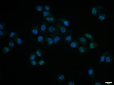
Hela cell; 4% Paraformaldehyde-fixed; Triton X-100 at room temperature for 20 min; Blocking buffer (normal goat serum, SLC0005) at 37°C for 20 min; Antibody incubation with (Phospho-PKR (Thr451)) polyclonal Antibody, Unconjugated (SL3336R) 1:100, 90 minutes at 37°C; followed by a conjugated Goat Anti-Rabbit IgG antibody at 37°C for 90 minutes, DAPI (blue, C02-04002) was used to stain the cell nuclei.
|