Home > Product > Antibody > Rabbit Anti-RALDH2 antibody
AL1A2_HUMAN; Aldehyde dehydrogenase family 1 member A2; ALDH1A2 aldehyde dehydrogenase 1 family, member A2; ALDH1A2; Aldh1a7; AV116159; MGC26444; RALDH 2; RALDH(II); RALDH2 T; Retinal dehydrogenase 2; Retinaldehyde dehydrogenase 2; Retinaldehy
Cat:
SL3676R
Species Reactivity:
Human,Mouse,Rat,(predicted: Dog,Pig,Cow,Horse,Rabbit,)
Immunogen:
KLH conjugated synthetic peptide derived from human RALDH2:441-518/518
Format:
Liquid
Storage instructions:
Shipped at 4℃. Store at -20 °C for one year. Avoid repeated freeze/thaw cycles.
Concentration:
1mg/ml
Clonality:
Polyclonal
Isotype:
IgG
Applications:
WB=1:500-2000ELISA=1:5000-10000IHC-P=1:100-500IHC-F=1:100-500IF=1:100-500(Paraffin sections need to do antigen repair)not yet tested in other applications.optimal dilutions/concentrations should be determined by the end user.
Host:
Rabbit
Product Overview:
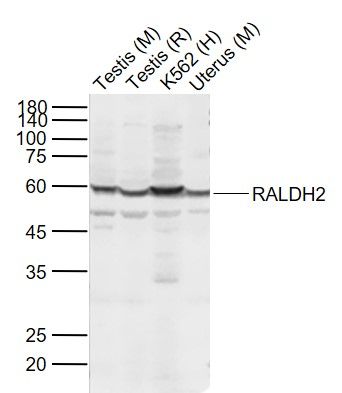
Sample: Lane 1: Testis (Mouse) Lysate at 40 ugLane 2: Testis (Rat) Lysate at 40 ugLane 3: K562 (Human) Cell Lysate at 30 ugLane 4: Uterus (Mouse) Lysate at 40 ugPrimary: Anti-RALDH2 (SL3676R) at 1/1000 dilutionSecondary: IRDye800CW Goat Anti-Rabbit IgG at 1/20000 dilutionPredicted band size: 56 kDObserved band size: 60 kDParaformaldehyde-fixed, paraffin embedded (Human kidney); Antigen retrieval by boiling in sodium citrate buffer (pH6.0) for 15min; Block endogenous peroxidase by 3% hydrogen peroxide for 20 minutes; Blocking buffer (normal goat serum) at 37°C for 30min; Antibody incubation with (RALDH2) Polyclonal Antibody, Unconjugated (SL3676R) at 1:200 overnight at 4°C, followed by operating according to SP Kit(Rabbit) (sp-0023) instructionsand DAB staining.Tissue/cell: rat brain tissue; 4% Paraformaldehyde-fixed and paraffin-embedded; Antigen retrieval: citrate buffer ( 0.01M, pH 6.0 ), Boiling bathing for 15min; Block endogenous peroxidase by 3% Hydrogen peroxide for 30min; Blocking buffer (normal goat serum,SLC0005) at 37℃ for 20 min; Incubation: Anti-RALDH2 Polyclonal Antibody, Unconjugated(SL3676R) 1:200, overnight at 4°C, followed by conjugation to the secondary antibody(SP-0023) and DAB(SLC0010) stainingTissue/cell: mouse embryo tissue;4% Paraformaldehyde-fixed and paraffin-embedded; Antigen retrieval: citrate buffer ( 0.01M, pH 6.0 ), Boiling bathing for 15min; Blocking buffer (normal goat serum,SLC0005) at 37℃ for 20 min; Incubation: Anti-RALDH2 Polyclonal Antibody, Unconjugated(SL3676R) 1:200, overnight at 4°C; The secondary antibody was Goat Anti-Rabbit IgG, Cy3 conjugated(SL0295G-Cy3)used at 1:200 dilution for 40 minutes at 37°C. DAPI(5ug/ml,blue,SLC0033) was used to stain the cell nucleiTissue/cell: rat brain tissue;4% Paraformaldehyde-fixed and paraffin-embedded; Antigen retrieval: citrate buffer ( 0.01M, pH 6.0 ), Boiling bathing for 15min; Blocking buffer (normal goat serum,SLC0005) at 37℃ for 20 min; Incubation: Anti-RALDH2 Polyclonal Antibody, Unconjugated(SL3676R) 1:200, overnight at 4°C; The secondary antibody was Goat Anti-Rabbit IgG, Cy3 conjugated(SL0295G-Cy3)used at 1:200 dilution for 40 minutes at 37°C. DAPI(5ug/ml,blue,SLC0033) was used to stain the cell nuclei
More
Unit:
Price: $
Product PDFs
Datasheet:


ALDH1A2 belongs to the aldehyde dehydrogenase family of proteins. It is an enzyme that catalyzes the synthesis of retinoic acid (RA) from retinaldehyde. Retinoic acid, the active derivative of vitamin A (retinol), is a hormonal signaling molecule that functions in developing and adult tissues. The studies of a similar mouse gene suggest that this enzyme and the cytochrome CYP26A1, concurrently establish local embryonic retinoic acid levels which facilitate posterior organ development and prevent spina bifida.

Subunit:
Homotetramer.

Subcellular Location:
Cytoplasm.

Similarity:
Belongs to the aldehyde dehydrogenase family.

SWISS:
O94788

Gene ID:
8854

Database links:

Entrez Gene: 8854 Human

Entrez Gene: 19378 Mouse

Entrez Gene: 116676 Rat

Omim: 603687 Human

SwissProt: O94788 Human

SwissProt: Q62148 Mouse

SwissProt: Q63639 Rat

Unigene: 643455 Human

Unigene: 42016 Mouse

Unigene: 10514 Rat


Picture

Sample:
Lane 1: Testis (Mouse) Lysate at 40 ug
Lane 2: Testis (Rat) Lysate at 40 ug
Lane 3: K562 (Human) Cell Lysate at 30 ug
Lane 4: Uterus (Mouse) Lysate at 40 ug
Primary: Anti-RALDH2 (SL3676R) at 1/1000 dilution
Secondary: IRDye800CW Goat Anti-Rabbit IgG at 1/20000 dilution
Predicted band size: 56 kD
Observed band size: 60 kD
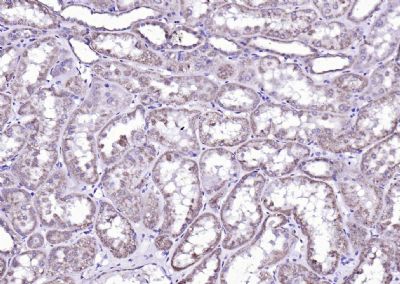
Paraformaldehyde-fixed, paraffin embedded (Human kidney); Antigen retrieval by boiling in sodium citrate buffer (pH6.0) for 15min; Block endogenous peroxidase by 3% hydrogen peroxide for 20 minutes; Blocking buffer (normal goat serum) at 37°C for 30min; Antibody incubation with (RALDH2) Polyclonal Antibody, Unconjugated (SL3676R) at 1:200 overnight at 4°C, followed by operating according to SP Kit(Rabbit) (sp-0023) instructionsand DAB staining.
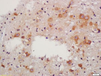
Tissue/cell: rat brain tissue; 4% Paraformaldehyde-fixed and paraffin-embedded;
Antigen retrieval: citrate buffer ( 0.01M, pH 6.0 ), Boiling bathing for 15min; Block endogenous peroxidase by 3% Hydrogen peroxide for 30min; Blocking buffer (normal goat serum,SLC0005) at 37℃ for 20 min;
Incubation: Anti-RALDH2 Polyclonal Antibody, Unconjugated(SL3676R) 1:200, overnight at 4°C, followed by conjugation to the secondary antibody(SP-0023) and DAB(SLC0010) staining
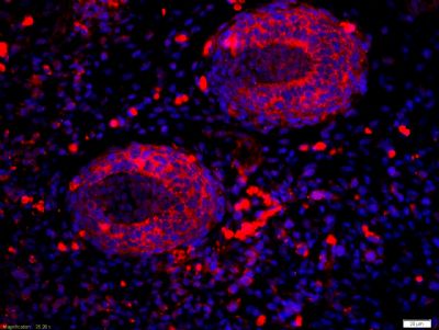
Tissue/cell: mouse embryo tissue;4% Paraformaldehyde-fixed and paraffin-embedded;
Antigen retrieval: citrate buffer ( 0.01M, pH 6.0 ), Boiling bathing for 15min; Blocking buffer (normal goat serum,SLC0005) at 37℃ for 20 min;
Incubation: Anti-RALDH2 Polyclonal Antibody, Unconjugated(SL3676R) 1:200, overnight at 4°C; The secondary antibody was Goat Anti-Rabbit IgG, Cy3 conjugated(SL0295G-Cy3)used at 1:200 dilution for 40 minutes at 37°C. DAPI(5ug/ml,blue,SLC0033) was used to stain the cell nuclei
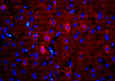
Tissue/cell: rat brain tissue;4% Paraformaldehyde-fixed and paraffin-embedded;
Antigen retrieval: citrate buffer ( 0.01M, pH 6.0 ), Boiling bathing for 15min; Blocking buffer (normal goat serum,SLC0005) at 37℃ for 20 min;
Incubation: Anti-RALDH2 Polyclonal Antibody, Unconjugated(SL3676R) 1:200, overnight at 4°C; The secondary antibody was Goat Anti-Rabbit IgG, Cy3 conjugated(SL0295G-Cy3)used at 1:200 dilution for 40 minutes at 37°C. DAPI(5ug/ml,blue,SLC0033) was used to stain the cell nuclei
Product Feedback Wall
Message :
Your Email :
Copyright © 2007-2018 Sunlong Medical All Rights Reserved.