Histones are basic nuclear proteins that are responsible for the nucleosome structure of the chromosomal fiber in eukaryotes. Two molecules of each of the four core histones (H2A, H2B, H3, and H4) form an octamer, around which approximately 146 bp of DNA is wrapped in repeating units, called nucleosomes. The linker histone, H1, interacts with linker DNA between nucleosomes and functions in the compaction of chromatin into higher order structures. This gene is intronless and encodes a member of the histone H2A family. Transcripts from this gene lack polyA tails but instead contain a palindromic termination element. This gene is found in the small histone gene cluster on chromosome 6p22-p21.3. [provided by RefSeq, Jul 2008].
Function:
Core component of nucleosome. Nucleosomes wrap and compact DNA into chromatin, limiting DNA accessibility to the cellular machineries which require DNA as a template. Histones thereby play a central role in transcription regulation, DNA repair, DNA replication and chromosomal stability. DNA accessibility is regulated via a complex set of post-translational modifications of histones, also called histone code, and nucleosome remodeling.
Subunit:
The nucleosome is a histone octamer containing two molecules each of H2A, H2B, H3 and H4 assembled in one H3-H4 heterotetramer and two H2A-H2B heterodimers. The octamer wraps approximately 147 bp of DNA.
Subcellular Location:
Nucleus. Chromosome.
Post-translational modifications:
The chromatin-associated form is phosphorylated on Thr-121 during mitosis (Probable).
Deiminated on Arg-4 in granulocytes upon calcium entry.
Monoubiquitination of Lys-120 by RING1 and RNF2/RING2 complex gives a specific tag for epigenetic transcriptional repression and participates in X chromosome inactivation of female mammals. It is involved in the initiation of both imprinted and random X inactivation. Ubiquitinated H2A is enriched in inactive X chromosome chromatin. Ubiquitination of H2A functions downstream of methylation of 'Lys-27' of histone H3. Monoubiquitination of Lys-120 by RNF2/RING2 can also be induced by ultraviolet and may be involved in DNA repair. Following DNA double-strand breaks (DSBs), it is ubiquitinated through 'Lys-63' linkage of ubiquitin moieties by the E2 ligase UBE2N and the E3 ligases RNF8 and RNF168, leading to the recruitment of repair proteins to sites of DNA damage. Monoubiquitination and ionizing radiation-induced 'Lys-63'-linked ubiquitination are distinct events.
Phosphorylation on Ser-2 is enhanced during mitosis. Phosphorylation on Ser-2 by RPS6KA5/MSK1 directly represses transcription. Acetylation of H3 inhibits Ser-2 phosphorylation by RPS6KA5/MSK1.
Symmetric dimethylation on Arg-4 by the PRDM1/PRMT5 complex may play a crucial role in the germ-cell lineage.
Similarity:
Belongs to the histone H2A family.
SWISS:
Q16777
Gene ID:
3012
Database links:
Entrez Gene: 3012 Human
Entrez Gene: 317772 Human
Entrez Gene: 8335 Human
Entrez Gene: 8337 Human
Entrez Gene: 8338 Human
Entrez Gene: 319166 Mouse
Omim: 142720 Human
Omim: 602786 Human
Omim: 602797 Human
SwissProt: P04908 Human
SwissProt: P28001 Human
SwissProt: P35065 Human
SwissProt: Q93077 Human
SwissProt: Q99878 Human
SwissProt: P10812 Mouse
SwissProt: P22752 Mouse
Unigene: 121017 Human
Unigene: 248174 Human
Unigene: 408067 Human
Unigene: 417332 Human
Unigene: 434195 Human
Unigene: 261665 Mouse
| Picture |
Tissue/cell: mouse embryo tissue; 4% Paraformaldehyde-fixed and paraffin-embedded;
Antigen retrieval: citrate buffer ( 0.01M, pH 6.0 ), Boiling bathing for 15min; Block endogenous peroxidase by 3% Hydrogen peroxide for 30min; Blocking buffer (normal goat serum,SLC0005) at 37℃ for 20 min;
Incubation: Anti-Histone H2A Polyclonal Antibody, Unconjugated(SL3779R) 1:200, overnight at 4°C, followed by conjugation to the secondary antibody(SP-0023) and DAB(SLC0010) staining
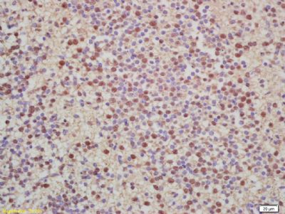
Tissue/cell: rat colon tissue; 4% Paraformaldehyde-fixed and paraffin-embedded;
Antigen retrieval: citrate buffer ( 0.01M, pH 6.0 ), Boiling bathing for 15min; Block endogenous peroxidase by 3% Hydrogen peroxide for 30min; Blocking buffer (normal goat serum,SLC0005) at 37℃ for 20 min;
Incubation: Anti-Histone H2A / H2A.1 Polyclonal Antibody, Unconjugated(SL3779R) 1:200, overnight at 4°C, followed by conjugation to the secondary antibody(SP-0023) and DAB(SLC0010) staining
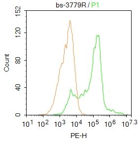
Blank control: HepG2.
Primary Antibody (green line): Rabbit Anti-Histone H2A antibody (SL3779R)
Dilution: 1μg /10^6 cells;
Isotype Control Antibody (orange line): Rabbit IgG .
Secondary Antibody : Goat anti-rabbit IgG-PE
Dilution: 1μg /test.
Protocol
The cells were fixed with 4% PFA (10min at room temperature)and then permeabilized with 90% ice-cold methanol for 20 min at-20℃. The cells were then incubated in 5%BSA to block non-specific protein-protein interactions for 30 min at at room temperature .Cells stained with Primary Antibody for 30 min at room temperature. The secondary antibody used for 40 min at room temperature. Acquisition of 20,000 events was performed.
|
|
|