Home > Product > Antibody > Rabbit Anti-PABP antibody
PAB 1; PAB1; PABP 1; PABP1; PABP-1; PABPC 1; PABPC1; PABPC2; PABPL1v Poly A binding protein 1; Poly A binding protein cytoplasmic 1; Polyadenylate binding protein 1.
Cat:
SL3833R
Species Reactivity:
Human,Mouse,Rat,(predicted: Pig,Horse,Rabbit,)
Immunogen:
KLH conjugated synthetic peptide derived from human PABP:101-200/636
Format:
Liquid
Storage instructions:
Shipped at 4℃. Store at -20 °C for one year. Avoid repeated freeze/thaw cycles.
Concentration:
1mg/ml
Clonality:
Polyclonal
Isotype:
IgG
Applications:
WB=1:500-2000ELISA=1:5000-10000IHC-P=1:100-500IHC-F=1:100-500Flow-Cyt=1ug/TestIF=1:100-500(Paraffin sections need to do antigen repair)not yet tested in other applications.optimal dilutions/concentrations should be determined by the end user.
Host:
Rabbit
Product Overview:
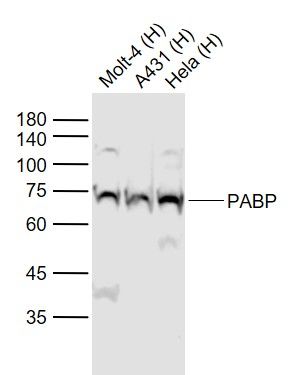
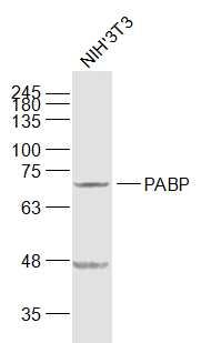
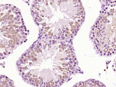
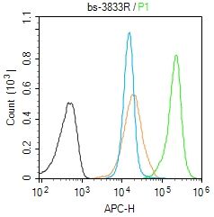
Sample: Lane 1: Molt-4 (Human) Cell Lysate at 30 ugLane 2: A431 (Human) Cell Lysate at 30 ugLane 3: Hela (Human) Cell Lysate at 30 ugPrimary: Anti-PABP (SL3833R) at 1/1000 dilutionSecondary: IRDye800CW Goat Anti-Rabbit IgG at 1/20000 dilutionPredicted band size: 70 kDObserved band size: 72 kDSample: NIH/3T3(Mouse) Cell Lysate at 30 ugPrimary: Anti-PABP (SL3833R) at 1/1000 dilutionSecondary: IRDye800CW Goat Anti-Rabbit IgG at 1/20000 dilutionPredicted band size: 70 kDObserved band size: 70 kD Paraformaldehyde-fixed, paraffin embedded (Mouse testis); Antigen retrieval by boiling in sodium citrate buffer (pH6.0) for 15min; Block endogenous peroxidase by 3% hydrogen peroxide for 20 minutes; Blocking buffer (normal goat serum) at 37°C for 30min; Antibody incubation with (PABP) Polyclonal Antibody, Unconjugated (SL3833R) at 1:400 overnight at 4°C, followed by operating according to SP Kit(Rabbit) (sp-0023) instructionsand DAB staining.Blank control (Black line):Molt4 (Black). Primary Antibody (green line): Rabbit Anti-PABP antibody (SL3833R) Dilution: 1μg /10^6 cells; Isotype Control Antibody (orange line): Rabbit IgG . Secondary Antibody (white blue line): Goat anti-rabbit IgG-AF647Dilution: 1μg /test. ProtocolThe cells were fixed with 4% PFA (10min at room temperature)and then permeabilized with 90% ice-cold methanol for 20 min at room temperature. The cells were then incubated in 5%BSA to block non-specific protein-protein interactions for 30 min at room temperature .Cells stained with Primary Antibody for 30 min at room temperature. The secondary antibody used for 40 min at room temperature. Acquisition of 20,000 events was performed.
More
Unit:
Price: $
Product PDFs
Datasheet:


This gene encodes a poly(A) binding protein. The protein shuttles between the nucleus and cytoplasm and binds to the 3' poly(A) tail of eukaryotic messenger RNAs via RNA-recognition motifs. The binding of this protein to poly(A) promotes ribosome recruitment and translation initiation; it is also required for poly(A) shortening which is the first step in mRNA decay. The gene is part of a small gene family including three protein-coding genes and several pseudogenes.[provided by RefSeq, Aug 2010].

Function:
Binds the poly(A) tail of mRNA. May be involved in cytoplasmic regulatory processes of mRNA metabolism such as pre-mRNA splicing. Its function in translational initiation regulation can either be enhanced by PAIP1 or repressed by PAIP2. Can probably bind to cytoplasmic RNA sequences other than poly(A) in vivo. Involved in translationally coupled mRNA turnover. Implicated with other RNA-binding proteins in the cytoplasmic deadenylation/translational and decay interplay of the FOS mRNA mediated by the major coding-region determinant of instability (mCRD) domain. Involved in regulation of nonsense-mediated decay (NMD) of mRNAs containing premature stop codons; for the recognition of premature termination codons (PTC) and initiation of NMD a competitive interaction between UPF1 and PABPC1 with the ribosome-bound release factors is proposed.

Subunit:
Component of a multi subunit autoregulatory ribonucleoprotein complex (ARC), at least composed of IGF2BP1, PABPC1 and CSDE1. Interacts with IGF2BP1.

Subcellular Location:
Cytoplasm. Nucleus.

Tissue Specificity:
Ubiquitous.

Post-translational modifications:
Phosphorylated by MAPKAPK2.
Methylated by CARM1. Arg-493 is dimethylated, probably to asymmetric dimethylarginine.

Similarity:
Belongs to the polyadenylate-binding protein type-1 family.
Contains 1 PABC domain.
Contains 4 RRM (RNA recognition motif) domains.

SWISS:
P11940

Gene ID:
26986

Database links:

Entrez Gene: 26986 Human

Entrez Gene: 18458 Mouse

Entrez Gene: 171350 Rat

Omim: 604679 Human

SwissProt: P11940 Human

SwissProt: P29341 Mouse

SwissProt: Q9EPH8 Rat

Unigene: 387804 Human

Unigene: 371570 Mouse

Unigene: 127813 Rat


Picture

Sample:
Lane 1: Molt-4 (Human) Cell Lysate at 30 ug
Lane 2: A431 (Human) Cell Lysate at 30 ug
Lane 3: Hela (Human) Cell Lysate at 30 ug
Primary: Anti-PABP (SL3833R) at 1/1000 dilution
Secondary: IRDye800CW Goat Anti-Rabbit IgG at 1/20000 dilution
Predicted band size: 70 kD
Observed band size: 72 kD
Sample:
NIH/3T3(Mouse) Cell Lysate at 30 ug
Primary: Anti-PABP (SL3833R) at 1/1000 dilution
Secondary: IRDye800CW Goat Anti-Rabbit IgG at 1/20000 dilution
Predicted band size: 70 kD
Observed band size: 70 kD
Paraformaldehyde-fixed, paraffin embedded (Mouse testis); Antigen retrieval by boiling in sodium citrate buffer (pH6.0) for 15min; Block endogenous peroxidase by 3% hydrogen peroxide for 20 minutes; Blocking buffer (normal goat serum) at 37°C for 30min; Antibody incubation with (PABP) Polyclonal Antibody, Unconjugated (SL3833R) at 1:400 overnight at 4°C, followed by operating according to SP Kit(Rabbit) (sp-0023) instructionsand DAB staining.
Blank control (Black line):Molt4 (Black).
Primary Antibody (green line): Rabbit Anti-PABP antibody (SL3833R)
Dilution: 1μg /10^6 cells;
Isotype Control Antibody (orange line): Rabbit IgG .
Secondary Antibody (white blue line): Goat anti-rabbit IgG-AF647
Dilution: 1μg /test.
Protocol
The cells were fixed with 4% PFA (10min at room temperature)and then permeabilized with 90% ice-cold methanol for 20 min at room temperature. The cells were then incubated in 5%BSA to block non-specific protein-protein interactions for 30 min at room temperature .Cells stained with Primary Antibody for 30 min at room temperature. The secondary antibody used for 40 min at room temperature. Acquisition of 20,000 events was performed.
Product Feedback Wall
Message :
Your Email :
Copyright © 2007-2018 Sunlong Medical All Rights Reserved.