CYP2R1 is a member of the cytochrome P450 superfamily of enzymes. The cytochrome P450 proteins are mono-oxygenases which catalyze many reactions involved in drug metabolism and synthesis of cholesterol, steroids and other lipids. This enzyme is a microsomal vitamin D hydroxylase that converts vitamin D into the active ligand for the vitamin D receptor.
Defects in CYP2R1 are a cause of 25-hydroxyvitamin D(3) deficiency, also known as pseudovitamin D(3) deficiency rickets due to 25-hydroxylase deficiency. First described in patients who had rickets at a young age despite a history of adequate vitamin D intake. The patients sera had low calcium concentrations, low phosphate concentrations, elevated alkaline phosphatase activity and low levels of 25-hydroxyvitamin D.
Subunit:
Homodimer.
Subcellular Location:
Endoplasmic reticulum membrane; Peripheral membrane protein. Microsome membrane; Peripheral membrane protein.
Tissue Specificity:
Endoplasmic reticulum membrane; Peripheral membrane protein. Microsome membrane; Peripheral membrane protein.
Similarity:
Belongs to the cytochrome P450 family.
SWISS:
Q6VVX0
Gene ID:
120227
Database links:
Entrez Gene: 120227 Human
Entrez Gene: 244209 Mouse
Omim: 608713 Human
SwissProt: Q6VVX0 Human
SwissProt: Q6VVW9 Mouse
Unigene: 371427 Human
Unigene: 108037 Mouse
| Picture |
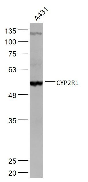
Sample:
A431(Human) Cell Lysate at 30 ug
Primary: Anti-CYP2R1 (SL3900R) at 1/1000 dilution
Secondary: IRDye800CW Goat Anti-Rabbit IgG at 1/20000 dilution
Predicted band size: 55 kD
Observed band size: 55 kD
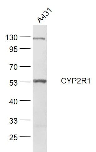
Sample:
A431(Human) Cell Lysate at 30 ug
Primary: Anti- CYP2R1 (SL3900R) at 1/1000 dilution
Secondary: IRDye800CW Goat Anti-Rabbit IgG at 1/20000 dilution
Predicted band size: 55 kD
Observed band size: 53 kD
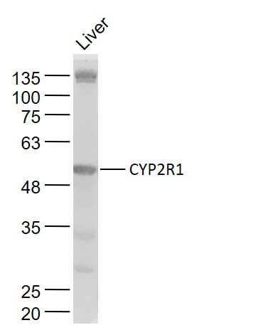
Sample:
Liver(Human) Cell Lysate at 40 ug
Primary: Anti-CYP2R1 (SL3900R) at 1/1000 dilution
Secondary: IRDye800CW Goat Anti-Rabbit IgG at 1/20000 dilution
Predicted band size: 55 kD
Observed band size: 55 kD
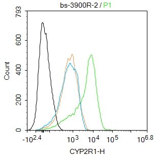
Blank control:Jurkat.
Primary Antibody (green line): Rabbit Anti-CYP2R1 antibody (SL3900R)
Dilution: 2ug/Test;
Secondary Antibody : Goat anti-rabbit IgG-FITC
Dilution: 0.5ug/Test.
Protocol
The cells were fixed with 4% PFA (10min at room temperature)and then permeabilized with 0.1% PBST for 20 min at room temperature.The cells were then incubated in 5%BSA to block non-specific protein-protein interactions for 30 min at room temperature.Cells stained with Primary Antibody for 30 min at room temperature. The secondary antibody used for 40 min at room temperature. Acquisition of 20,000 events was performed.
|
|
|