Home > Product > Antibody > Rabbit Anti-TXNIP antibody
;THIF; thioredoxin binding protein 2; thioredoxin interacting protein; Thioredoxin-binding protein 2; Thioredoxin-interacting protein; upregulated by 1,25-dihydroxyvitamin D-3; VDUP1; Vitamin D3 up-regulated protein 1; TXNIP_HUMAN.
Cat:
SL3897R
Species Reactivity:
Human,Rat,(predicted: Mouse,Dog,Pig,Rabbit,)
Immunogen:
KLH conjugated synthetic peptide derived from human TXNIP:101-200/391
Format:
Liquid
Storage instructions:
Shipped at 4℃. Store at -20 °C for one year. Avoid repeated freeze/thaw cycles.
Concentration:
1mg/ml
Clonality:
Polyclonal
Isotype:
IgG
Applications:
ELISA=1:5000-10000IHC-P=1:100-500IHC-F=1:100-500Flow-Cyt=1ug/testIF=1:100-500(Paraffin sections need to do antigen repair)not yet tested in other applications.optimal dilutions/concentrations should be determined by the end user.
Host:
Rabbit
Product Overview:
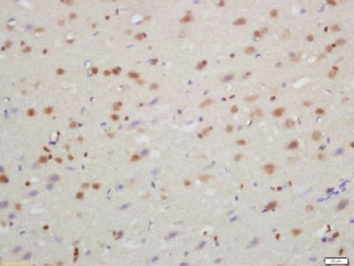
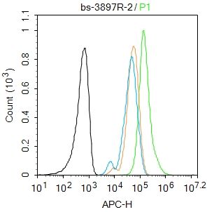
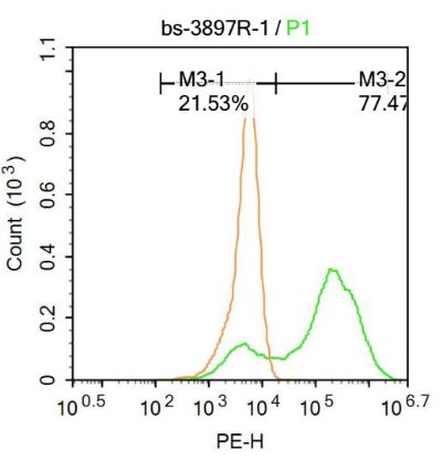
Tissue/cell: rat brain tissue; 4% Paraformaldehyde-fixed and paraffin-embedded; Antigen retrieval: citrate buffer ( 0.01M, pH 6.0 ), Boiling bathing for 15min; Block endogenous peroxidase by 3% Hydrogen peroxide for 30min; Blocking buffer (normal goat serum,SLC0005) at 37℃ for 20 min; Incubation: Anti-TXNIP Polyclonal Antibody, Unconjugated(SL3897R) 1:200, overnight at 4°C, followed by conjugation to the secondary antibody(SP-0023) and DAB(SLC0010) stainingBlank control: U937. Primary Antibody (green line): Rabbit Anti-TXNIP antibody (SL3897R) Dilution: 2μg /10^6 cells; Isotype Control Antibody (orange line): Rabbit IgG . Secondary Antibody : Goat anti-rabbit IgG-AF647Dilution: 1μg /test. ProtocolThe cells were fixed with 4% PFA (10min at room temperature)and then permeabilized with 90% ice-cold methanol for 20 min at-20℃.The cells were then incubated in 5%BSA to block non-specific protein-protein interactions for 30 min at at room temperature .Cells stained with Primary Antibody for 30 min at room temperature. The secondary antibody used for 40 min at room temperature. Acquisition of 20,000 events was performed.U-937 cells were fixed with 4% PFA for 10min at room temperature,permeabilized with 90% ice-cold methanol for 20 min at room temperature, and incubated in 5% BSA blocking buffer for 30 min at room temperature. Cells were then stained with TXNIP Antibody(SL3897R)at 1:100 dilution in blocking buffer and incubated for 30 min at room temperature, washed twice with 2%BSA in PBS, followed by secondary antibody incubation for 40 min at room temperature. Acquisitions of 20,000 events were performed. Cells stained with primary antibody (green), and isotype control (orange).
More
Unit:
Price: $
Product PDFs
Datasheet:


TXNIP may act as an oxidative stress mediator by inhibiting thioredoxin activity or by limiting its bioavailability. It interacts with COPS5 and restores COPS5-induced suppression of CDKN1B stability, blocking the COPS5-mediated translocation of CDKN1B from the nucleus to the cytoplasm. It functions as a transcriptional repressor, possibly by acting as a bridge molecule between transcription factors and corepressor complexes, and over-expression will induce G0/G1 cell cycle arrest. It is required for the maturation of natural killer cells (from SwissProt).

Function:
May act as an oxidative stress mediator by inhibiting thioredoxin activity or by limiting its bioavailability. Interacts with COPS5 and restores COPS5-induced suppression of CDKN1B stability, blocking the COPS5-mediated translocation of CDKN1B from the nucleus to the cytoplasm. Functions as a transcriptional repressor, possibly by acting as a bridge molecule between transcription factors and corepressor complexes, and over-expression will induce G0/G1 cell cycle arrest. Required for the maturation of natural killer cells. Acts as a suppressor of tumor cell growth. Inhibits the proteasomal degradation of DDIT4, and thereby contributes to the inhibition of the mammalian target of rapamycin complex 1 (mTORC1).

Subunit:
Interacts with TXN/thioredoxin through its redox-active site. Interacts with transcriptional repressors ZBTB16, ZBTB32 and HDAC1. Interacts (via SLCterminus) with ITCH (via WW domains). Interacts with DDIT4.

Subcellular Location:
Cytoplasm

Post-translational modifications:
Ubiquitinated; undergoes polyubiquitination catalyzed by ITCH resulting in proteasomal degradation.

Similarity:
Belongs to the arrestin family.

SWISS:
Q9H3M7

Gene ID:
10628

Database links:
UniProtKB/Swiss-Prot: Q9H3M7.1

    硫氧还蛋白结合蛋白-2 (TRX-binding protein-2,TBP-2)又称:维生素D3上调蛋白1(Vitamin D3 up-regulated protein 1,VDUP1)或称硫氧还蛋白相互作用蛋白(Thioredoxin-interacting protein,Txnip)。
    TBP-2与硫氧还蛋白(thioredoxin,TRX)的活性部位结合,抑制其活性,除了参与多种氧化应激反应,还具有多种生物学功能。
    有学者认为:TBP-2与肿瘤密切相关,有些肿瘤组织中,TBP-2表达降低或缺失。TBP-2表达增高时,细胞周期停滞,肿瘤细胞繁殖受到抑制。目前已成为肿瘤研究的重要靶点。 Picture

Tissue/cell: rat brain tissue; 4% Paraformaldehyde-fixed and paraffin-embedded;
Antigen retrieval: citrate buffer ( 0.01M, pH 6.0 ), Boiling bathing for 15min; Block endogenous peroxidase by 3% Hydrogen peroxide for 30min; Blocking buffer (normal goat serum,SLC0005) at 37℃ for 20 min;
Incubation: Anti-TXNIP Polyclonal Antibody, Unconjugated(SL3897R) 1:200, overnight at 4°C, followed by conjugation to the secondary antibody(SP-0023) and DAB(SLC0010) staining
Blank control: U937.
Primary Antibody (green line): Rabbit Anti-TXNIP antibody (SL3897R)
Dilution: 2μg /10^6 cells;
Isotype Control Antibody (orange line): Rabbit IgG .
Secondary Antibody : Goat anti-rabbit IgG-AF647
Dilution: 1μg /test.
Protocol
The cells were fixed with 4% PFA (10min at room temperature)and then permeabilized with 90% ice-cold methanol for 20 min at-20℃.The cells were then incubated in 5%BSA to block non-specific protein-protein interactions for 30 min at at room temperature .Cells stained with Primary Antibody for 30 min at room temperature. The secondary antibody used for 40 min at room temperature. Acquisition of 20,000 events was performed.
U-937 cells were fixed with 4% PFA for 10min at room temperature,permeabilized with 90% ice-cold methanol for 20 min at room temperature, and incubated in 5% BSA blocking buffer for 30 min at room temperature. Cells were then stained with TXNIP Antibody(SL3897R)at 1:100 dilution in blocking buffer and incubated for 30 min at room temperature, washed twice with 2%BSA in PBS, followed by secondary antibody incubation for 40 min at room temperature. Acquisitions of 20,000 events were performed. Cells stained with primary antibody (green), and isotype control (orange).
Product Feedback Wall
Message :
Your Email :
Copyright © 2007-2018 Sunlong Medical All Rights Reserved.