Guanine nucleotide-binding proteins (G proteins) are involved as modulators or transducers in various transmembrane signaling systems. The Gs protein is involved in hormonal regulation of adenylate cyclase: it activates the cyclase in response to beta-adrenergic stimuli.
Function:
Guanine nucleotide-binding proteins (G proteins) are involved as modulators or transducers in various transmembrane signaling systems. The G(s) protein is involved in hormonal regulation of adenylate cyclase: it activates the cyclase in response to beta-adrenergic stimuli. XLas isoforms interact with the same set of receptors as Gnas isoforms (By similarity).
Subunit:
G proteins are composed of 3 units; alpha, beta and gamma. The alpha chain contains the guanine nucleotide binding site. Interacts through its N-terminal region with ALEX which is produced from the same locus in a different open reading frame. This interaction may inhibit its adenylyl cyclase-stimulating activity (By similarity).
Subcellular Location:
Cell membrane; Peripheral membrane protein.
DISEASE:
Defects in GNAS are the cause of GNAS hyperfunction (GNASHYP) [MIM:13964]. This condition is characterized by increased trauma-related bleeding tendency, prolonged bleeding time, brachydactyly and mental retardation. Both the XLas isoforms and the ALEX protein are mutated which strongly reduces the interaction between them and this may allow unimpeded activation of the XLas isoforms.
Defects in GNAS are a cause of ACTH-independent macronodular adrenal hyperplasia (AIMAH) [MIM:219080]; also known as adrenal Cushing syndrome due to AIMAH. A rare adrenal defect characterized by multiple, bilateral, non-pigmented, benign, adrenocortical nodules. It results in excessive production of cortisol leading to ACTH-independent Cushing syndrome. Clinical manifestations of Cushing syndrome include facial and trunkal obesity, abdominal striae, muscular weakness, osteoporosis, arterial hypertension, diabetes.
Genetic variations in GNAS are the cause of pseudohypoparathyroidism type 1B (PHP1B) [MIM:603233]. PHP1B is characterized by parathyroid hormone (PTH)-resistant hypocalcemia and hyperphosphatemia. Patients affected with PHP1B have normal activity of the product of GNAS, lack developmental defects characteristic of AHO, and typically show no other endocrine abnormalities besides resistance to PTH. Note=Most affected individuals have defects in methylation of the gene. In some cases microdeletions involving the STX16 appear to cause loss of methylation at exon A/B of GNAS, resulting in PHP1B. Paternal uniparental isodisomy have also been observed.
Defects in GNAS are the cause of pseudohypoparathyroidism type 1C (PHP1C) [MIM:612462]. It is a disorder characterized by end-organ resistance to parathyroid hormone, hypocalcemia and hyperphosphatemia. It is commonly associated with Albright hereditary osteodystrophy whose features are short stature, obesity, round facies, short metacarpals and ectopic calcification.
Similarity:
Belongs to the G-alpha family. G(s) subfamily. membrane protein.
SWISS:
Q5JWF2
Gene ID:
2778
Database links:
Entrez Gene: 281793 Cow
Entrez Gene: 2778 Human
Entrez Gene: 14683 Mouse
Entrez Gene: 100049657 Pig
Entrez Gene: 24896 Rat
Omim: 13964 Human
SwissProt: P04896 Cow
SwissProt: P63091 Dog
SwissProt: P63092 Human
SwissProt: P84996 Human
SwissProt: Q5JWF2 Human
SwissProt: P63094 Mouse
SwissProt: Q6R0H7 Mouse
SwissProt: P29797 Pig
SwissProt: P63095 Rat
SwissProt: Q63803 Rat
| Picture |
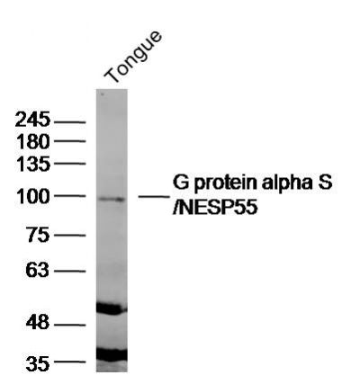
Sample: tongue(mouse) Lysate at 40 ug
Primary: Anti- G protein alpha S'NESP55 (SL3939R)at 1/300 dilution
Secondary: IRDye800CW Goat Anti-Rabbit IgG at 1/20000 dilution
Predicted band size: 111kD
Observed band size: 100 kD
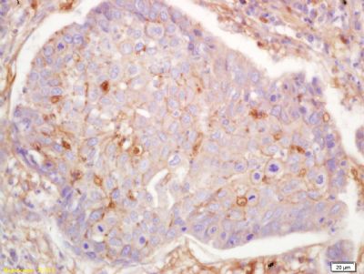
Tissue/cell: human lung carcinoma; 4% Paraformaldehyde-fixed and paraffin-embedded;
Antigen retrieval: citrate buffer ( 0.01M, pH 6.0 ), Boiling bathing for 15min; Block endogenous peroxidase by 3% Hydrogen peroxide for 30min; Blocking buffer (normal goat serum,SLC0005) at 37℃ for 20 min;
Incubation: Anti-G protein alpha S Polyclonal Antibody, Unconjugated(SL3939R) 1:200, overnight at 4°C, followed by conjugation to the secondary antibody(SP-0023) and DAB(SLC0010) staining
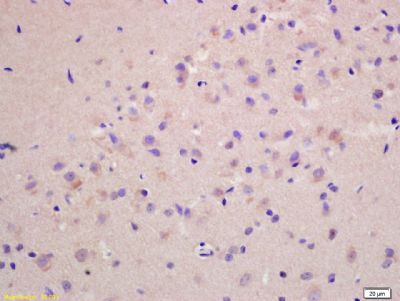
Tissue/cell: rat brain tissue; 4% Paraformaldehyde-fixed and paraffin-embedded;
Antigen retrieval: citrate buffer ( 0.01M, pH 6.0 ), Boiling bathing for 15min; Block endogenous peroxidase by 3% Hydrogen peroxide for 30min; Blocking buffer (normal goat serum,SLC0005) at 37℃ for 20 min;
Incubation: Anti-G protein alpha S Polyclonal Antibody, Unconjugated(SL3939R) 1:200, overnight at 4°C, followed by conjugation to the secondary antibody(SP-0023) and DAB(SLC0010) staining
|
|
|