Home > Product > Antibody > Rabbit Anti-SMURF2 antibody
hSMURF2; MGC138150; Smad specific E3 ubiquitin ligase 2; SMAD specific E3 ubiquitin protein ligase 2; Smad ubiquitination regulatory factor 2; Ubiquitin protein ligase SMURF2; DKFZp686F0270; MGC138150; E3 ubiquitin-protein ligase SMURF2; EC 6.3.2.; SMAD-s
Cat:
SL4056R
Species Reactivity:
Human,Mouse,(predicted: Rat,Chicken,Cow,Horse,)
Immunogen:
KLH conjugated synthetic peptide derived from human SMURF2:601-700/748
Format:
Liquid
Storage instructions:
Shipped at 4℃. Store at -20 °C for one year. Avoid repeated freeze/thaw cycles.
Concentration:
1mg/ml
Clonality:
Polyclonal
Isotype:
IgG
Applications:
WB=1:500-2000ELISA=1:5000-10000IHC-P=1:100-500IHC-F=1:100-500Flow-Cyt=3ug/testIF=1:100-500(Paraffin sections need to do antigen repair)not yet tested in other applications.optimal dilutions/concentrations should be determined by the end user.
Host:
Rabbit
Product Overview:
Sample: Placenta (Mouse) Lysate at 40 ugPrimary: Anti-SMURF2 (SL4056R) at 1/300 dilutionSecondary: IRDye800CW Goat Anti-Rabbit IgG at 1/20000 dilutionPredicted band size: 82 kDObserved band size: 82 kDParaformaldehyde-fixed, paraffin embedded (mouse brain tissue); Antigen retrieval by microwave in sodium citratebuffer (pH6.0) ; Block endogenous peroxidase by 3% hydrogen peroxide for 30 minutes; Blocking buffer (3%BSA) at RTfor 30min; Antibody incubation with (SMURF2) Polyclonal/MonoclonalAntibody, Unconjugated (SL4056R) at 1:400 overnight at 4℃,followed by conjugation to the secondary antibody (labeled with HRP)and DAB staining.Blank control:A431. Primary Antibody (green line): Rabbit Anti-SMURF2 antibody (SL4056R) Dilution: 3μg /10^6 cells; Isotype Control Antibody (orange line): Rabbit IgG . Secondary Antibody : Goat anti-rabbit IgG-AF647Dilution: 3μg /test. ProtocolThe cells were fixed with 4% PFA (10min at room temperature)and then permeabilized with 90% ice-cold methanol for 20 min at-20℃. The cells were then incubated in 5%BSA to block non-specific protein-protein interactions for 30 min at at room temperature .Cells stained with Primary Antibody for 30 min at room temperature. The secondary antibody used for 40 min at room temperature. Acquisition of 20,000 events was performed.
More
Unit:
Price: $
Product PDFs
Datasheet:


SMURF2, a 748-amino acid ubiquitin E3 ligase that is 83% identical to SMURF1, codes for a C2-WW-HECT domain ubiquitin ligase that associates constitutively with SMAD7. Binding to SMAD7 induces export of SMURF2 and recruitment to the activated transforming growth factor-beta receptor (TGFBR), where it causes receptor and SMAD7 degradation. A strong interaction of second and third SMURF2 WW domains has been identified with SMAD1, SMAD2, and SMAD3, but not SMAD4. Western blot analysis showed that SMURF2 selectively downregulates the transcription of SMAD2 and SMAD1, but not SMAD3. The nuclear SMURF2/phosphorylated SMAD2 interaction is requires TGFB1.

Function:
E3 ubiquitin-protein ligase which accepts ubiquitin from an E2 ubiquitin-conjugating enzyme in the form of a thioester and then directly transfers the ubiquitin to targeted substrates. Interacts with SMAD1 and SMAD7 in order to trigger their ubiquitination and proteasome-dependent degradation. In addition, interaction with SMAD7 activates autocatalytic degradation, which is prevented by interaction with SCYE1. Forms a stable complex with the TGF-beta receptor-mediated phosphorylated SMAD2 and SMAD3. In this way, SMAD2 may recruit substrates, such as SNON, for ubiquitin-mediated degradation. Enhances the inhibitory activity of SMAD7 and reduces the transcriptional activity of SMAD2. Coexpression of SMURF2 with SMAD1 results in considerable decrease in steady-state level of SMAD1 protein and a smaller decrease of SMAD2 level.

Subunit:
Interacts (via WW domains) with SMAD1. Interacts (via WW domains) with SMAD2 (via PY-motif). Interacts (via WW domains) with SMAD3 (via PY-motif). Interacts with SMAD6. Interacts with SMAD7 (via PY-motif) and TGFBR1; SMAD7 recruits SMURF2 to the TGF-beta receptor and regulates its degradation. Does not interact with SMAD4; SMAD4 lacks a PY-motif. Interacts with AIMP1. Interacts with STAMBP and RNF11. Interacts with NDFIP1 and NDFIP2 (Probable); this interaction activates the E3 ubiquitin-protein ligase.

Subcellular Location:
Nucleus. Cytoplasm. Cell membrane. Membrane raft. Cytoplasmic in the presence of SMAD7. Co-localizes with CAV1, SMAD7 and TGF-beta receptor in membrane rafts.

Tissue Specificity:
Widely expressed.

Post-translational modifications:
Auto-ubiquitinated and ubiquitinated in the presence of RNF11 and UBE2D1. Ubiquitinated by the SCF(FBXL15) complex, leading to its degradation by the proteasome.

Similarity:
Contains 1 C2 domain.
Contains 1 HECT (E6AP-type E3 ubiquitin-protein ligase) domain.
Contains 3 WW domains.

SWISS:
Q9HAU4

Gene ID:
64750

Database links:

Entrez Gene: 64750 Human

Entrez Gene: 66313 Mouse

Entrez Gene: 303614 Rat

Omim: 605532 Human

SwissProt: Q52LL1 Human

SwissProt: Q9HAU4 Human

SwissProt: A2A5Z6 Mouse

SwissProt: Q3TT87 Mouse

SwissProt: Q5IRE6 Mouse

Unigene: 340955 Mouse


Picture

Sample: Placenta (Mouse) Lysate at 40 ug
Primary: Anti-SMURF2 (SL4056R) at 1/300 dilution
Secondary: IRDye800CW Goat Anti-Rabbit IgG at 1/20000 dilution
Predicted band size: 82 kD
Observed band size: 82 kD
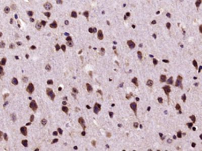
Paraformaldehyde-fixed, paraffin embedded (mouse brain tissue); Antigen retrieval by microwave in sodium citratebuffer (pH6.0) ; Block endogenous peroxidase by 3% hydrogen peroxide for 30 minutes; Blocking buffer (3%BSA) at RTfor 30min; Antibody incubation with (SMURF2) Polyclonal/MonoclonalAntibody, Unconjugated (SL4056R) at 1:400 overnight at 4℃,followed by conjugation to the secondary antibody (labeled with HRP)and DAB staining.
Blank control:A431.
Primary Antibody (green line): Rabbit Anti-SMURF2 antibody (SL4056R)
Dilution: 3μg /10^6 cells;
Isotype Control Antibody (orange line): Rabbit IgG .
Secondary Antibody : Goat anti-rabbit IgG-AF647
Dilution: 3μg /test.
Protocol
The cells were fixed with 4% PFA (10min at room temperature)and then permeabilized with 90% ice-cold methanol for 20 min at-20℃. The cells were then incubated in 5%BSA to block non-specific protein-protein interactions for 30 min at at room temperature .Cells stained with Primary Antibody for 30 min at room temperature. The secondary antibody used for 40 min at room temperature. Acquisition of 20,000 events was performed.
Product Feedback Wall
Message :
Your Email :
Copyright © 2007-2018 Sunlong Medical All Rights Reserved.