2 enoyl Coenzyme A (CoA) hydratase beta subunit; 3 ketoacyl Coenzyme A (CoA) thiolase of mitochondrial trifunctional protein beta subunit; 3 ketoacyl Coenzyme A thiolase; 3 ketoacyl Coenzyme A thiolase of mitochondrial trifunctional protein beta subunit;
KLH conjugated synthetic peptide derived from human HADHB:231-330/474
Format:
Liquid
Storage instructions:
Shipped at 4℃. Store at -20 °C for one year. Avoid repeated freeze/thaw cycles.
Concentration:
1mg/ml
Clonality:
Polyclonal
Isotype:
IgG
Applications:
WB=1:500-2000ELISA=1:5000-10000IHC-P=1:100-500IHC-F=1:100-500IF=1:100-500(Paraffin sections need to do antigen repair)not yet tested in other applications.optimal dilutions/concentrations should be determined by the end user.
Host:
Rabbit
Product Overview:
Sample: Kidney (Mouse) Lysate at 40 ugPrimary: Anti- HADHB (SL5065R) at 1/1000 dilutionSecondary: IRDye800CW Goat Anti-Rabbit IgG at 1/20000 dilutionPredicted band size: 47kDObserved band size: 47 kDSY5Y(Human) Cell Lysate at 30 ugPrimary: Anti-HADHB (SL5065R) at 1/1000 dilutionSecondary: IRDye800CW Goat Anti-Rabbit IgG at 1/20000 dilutionPredicted band size: 47 kDObserved band size: 47 kDSiha(Human) Cell Lysate at 30 ugPrimary: Anti-HADHB (SL5065R) at 1/1000 dilutionSecondary: IRDye800CW Goat Anti-Rabbit IgG at 1/20000 dilutionPredicted band size: 47 kDObserved band size: 47 kD
The HADHB gene encodes the beta subunit of the mitochondrial trifunctional protein, which catalyzes the last three steps of mitochondrial beta-oxidation of long chain fatty acids. The mitochondrial membrane-bound heterocomplex is composed of four alpha and four beta subunits, with the beta subunit catalyzing the 3-ketoacyl-CoA thiolase activity. Mutations in this gene result in trifunctional protein deficiency. The encoded protein can also bind RNA and decreases the stability of some mRNAs. The genes of the alpha and beta subunits of the mitochondrial trifunctional protein are located adjacent to each other in the human genome in a head-to-head orientation. Alternatively spliced transcript variants have been found; however, their full-length nature is not known.
Subunit: Octamer of 4 alpha (HADHA) and 4 beta (HADHB) subunits. Interacts with RSAD2/viperin.
DISEASE: Defects in HADHB are a cause of trifunctional protein deficiency (TFP deficiency) [MIM:609015]. The clinical manifestations are very variable and include hypoglycemia, cardiomyopathy and sudden death. Phenotypes with mainly hepatic and neuromyopathic involvement can also be distinguished. Biochemically, TFP deficiency is defined by the loss of all three enzyme activities of the TFP complex.
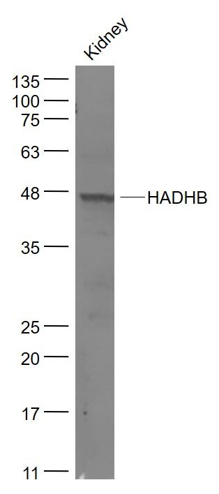
Sample:
Kidney (Mouse) Lysate at 40 ug
Primary: Anti- HADHB (SL5065R) at 1/1000 dilution
Secondary: IRDye800CW Goat Anti-Rabbit IgG at 1/20000 dilution
Predicted band size: 47kD
Observed band size: 47 kD
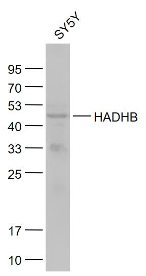
SY5Y(Human) Cell Lysate at 30 ug
Primary: Anti-HADHB (SL5065R) at 1/1000 dilution
Secondary: IRDye800CW Goat Anti-Rabbit IgG at 1/20000 dilution
Predicted band size: 47 kD
Observed band size: 47 kD
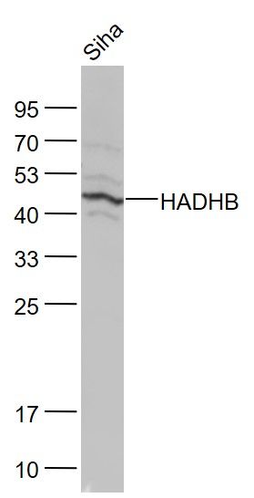
Siha(Human) Cell Lysate at 30 ug
Primary: Anti-HADHB (SL5065R) at 1/1000 dilution
Secondary: IRDye800CW Goat Anti-Rabbit IgG at 1/20000 dilution
Predicted band size: 47 kD
Observed band size: 47 kD