Home > Product > Antibody > Rabbit Anti-Lamin B Receptor antibody
LBR_HUMAN; LMN2R; PHA; DHCR 14B; DHCR14B antibody Integral nuclear envelope inner membrane protein; Lamin-B receptor; LBR; LMN 2R; LMN2R; MGC9041; PHA; PRO0650; DHCR14B; MGC9041.
Cat:
SL5081R
Species Reactivity:
Human,Mouse,(predicted: Rat,Cow,Horse,Rabbit,)
Immunogen:
KLH conjugated synthetic peptide derived from human Lamin B Receptor:1-100/615
Format:
Liquid
Storage instructions:
Shipped at 4℃. Store at -20 °C for one year. Avoid repeated freeze/thaw cycles.
Concentration:
1mg/ml
Clonality:
Polyclonal
Isotype:
IgG
Applications:
WB=1:500-2000ELISA=1:5000-10000Flow-Cyt=1ug/Testnot yet tested in other applications.optimal dilutions/concentrations should be determined by the end user.
Host:
Rabbit
Product Overview:
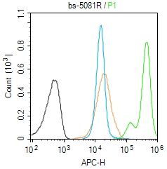
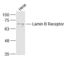
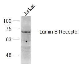
Sample: Hela(Human) Cell Lysate at 30 ugPrimary: Anti-Lamin B Receptor (SL5081R) at 1/1000 dilutionSecondary: IRDye800CW Goat Anti-Rabbit IgG at 1/20000 dilutionPredicted band size: 68 kDObserved band size: 68 kDSample: Jurkat(Human) Cell Lysate at 30 ugPrimary: Anti-Lamin B Receptor (SL5081R) at 1/1000 dilutionSecondary: IRDye800CW Goat Anti-Rabbit IgG at 1/20000 dilutionPredicted band size: 68 kDObserved band size: 68 kDBlank control (Black line):Molt4 (Black). Primary Antibody (green line): Rabbit Anti-Lamin B Receptor antibody (SL5801R) Dilution: 1μg /10^6 cells; Isotype Control Antibody (orange line): Rabbit IgG . Secondary Antibody (white blue line): Goat anti-rabbit IgG-AF647Dilution: 1μg /test. ProtocolThe cells were fixed with 4% PFA (10min at room temperature)and then permeabilized with 90% ice-cold methanol for 20 min at room temperature. The cells were then incubated in 5%BSA to block non-specific protein-protein interactions for 30 min at room temperature .Cells stained with Primary Antibody for 30 min at room temperature. The secondary antibody used for 40 min at room temperature. Acquisition of 20,000 events was performed.
More
Unit:
Price: $
Product PDFs
Datasheet:


Lamins are nuclear membrane proteins that serve to maintain specific cellular functions, such as DNA replication and chromatin organization. Lamin B receptor (LBR) is an integral protein of the nuclear envelope inner membrane. It is phosphorylated by CDC2 protein kinase in mitosis when the inner nuclear membrane breaks down into vesicles that dissociate from the lamina and the chromatin. It is phosphorylated by different protein kinases in interphase when the membrane is associated with these structures. The cleavage of lamins results in nuclear disregulation and cell death.

Function:
Anchors the lamina and the heterochromatin to the inner nuclear membrane.

Subunit:
Interacts directly with CBX5. Can interact with chromodomain proteins. Interacts directly with DNA. Interaction with DNA is sequence independent with higher affinity for supercoiled and relaxed circular DNA than linear DNA.

Subcellular Location:
Nucleus inner membrane; Multi-pass membrane protein.

Post-translational modifications:
Phosphorylated by CDK1 in mitosis when the inner nuclear membrane breaks down into vesicles that dissociate from the lamina and the chromatin. It is phosphorylated by different protein kinases in interphase when the membrane is associated with these structures. Phosphorylation of LBR and HP1 proteins may be responsible for some of the alterations in chromatin organization and nuclear structure which occur at various times during the cell cycle. Phosphorylated by SRPK1. In late anaphase LBR is dephosphorylated, probably by PP1 and/or PP2A, allowing reassociation with chromatin.

DISEASE:
Defects in LBR are a cause of Pelger-Huet anomaly (PHA) [MIM:169400]. PHA is an autosomal dominant inherited abnormality of neutrophils, characterized by reduced nuclear segmentation and an apparently looser chromatin structure. Heterozygotes show hypolobulated neutrophil nuclei with coarse chromatin. Presumed homozygous individuals have ovoid neutrophil nuclei, as well as varying degrees of developmental delay, epilepsy, and skeletal abnormalities.
[DISEASE] Defects in LBR are the cause of hydrops-ectopic calcification-moth-eaten skeletal dysplasia (HEM) [MIM:215140]; also known as Greenberg skeletal dysplasia. HEM is a rare autosomal recessive chondrodystrophy characterized by early in utero lethality and, therefore, considered to be nonviable. Affected fetuses typically present with fetal hydrops, short-limbed dwarfism, and a marked disorganization of chondro-osseous calcification and may present with polydactyly and additional nonskeletal malformations.

Similarity:
Belongs to the ERG4/ERG24 family.
Contains 1 Tudor domain.

SWISS:
Q14739

Gene ID:
3930

Database links:

Entrez Gene: 3930 Human

Entrez Gene: 89789 Rat

Omim: 600024 Human

SwissProt: Q14739 Human

SwissProt: O08984 Rat

Unigene: 435166 Human

Unigene: 6499 Rat


[DISEASE] Defects in LBR may be a cause of Reynolds syndrome (REYNS) [MIM:613471]. It is a syndrome specifically associating limited cutaneous systemic sclerosis and primary biliray cirrhosis. It is characterized by liver disease, telangiectasia, abrupt onset of digital paleness or cyanosis in response to cold exposure or stress (Raynaud phenomenon), and variable features of scleroderma. The liver disease is characterized by pruritis, jaundice, hepatomegaly, increased serum alkaline phosphatase and positive serum mitochondrial autoantibodies, all consistent with primary biliary cirrhosis. Picture

Sample:
Hela(Human) Cell Lysate at 30 ug
Primary: Anti-Lamin B Receptor (SL5081R) at 1/1000 dilution
Secondary: IRDye800CW Goat Anti-Rabbit IgG at 1/20000 dilution
Predicted band size: 68 kD
Observed band size: 68 kD
Sample:
Jurkat(Human) Cell Lysate at 30 ug
Primary: Anti-Lamin B Receptor (SL5081R) at 1/1000 dilution
Secondary: IRDye800CW Goat Anti-Rabbit IgG at 1/20000 dilution
Predicted band size: 68 kD
Observed band size: 68 kD
Blank control (Black line):Molt4 (Black).
Primary Antibody (green line): Rabbit Anti-Lamin B Receptor antibody (SL5801R)
Dilution: 1μg /10^6 cells;
Isotype Control Antibody (orange line): Rabbit IgG .
Secondary Antibody (white blue line): Goat anti-rabbit IgG-AF647
Dilution: 1μg /test.
Protocol
The cells were fixed with 4% PFA (10min at room temperature)and then permeabilized with 90% ice-cold methanol for 20 min at room temperature. The cells were then incubated in 5%BSA to block non-specific protein-protein interactions for 30 min at room temperature .Cells stained with Primary Antibody for 30 min at room temperature. The secondary antibody used for 40 min at room temperature. Acquisition of 20,000 events was performed.
Product Feedback Wall
Message :
Your Email :
Copyright © 2007-2018 Sunlong Medical All Rights Reserved.