Involved in the homologous recombination repair (HRR) pathway of double-stranded DNA, thought to repair chromosomal fragmentation, translocations and deletions. Plays a role in regulating mitochondrial DNA copy number under conditions of oxidative stress in the presence of RAD51 and RAD51C.
Function:
Involved in the homologous recombination repair (HRR) pathway of double-stranded DNA, thought to repair chromosomal fragmentation, translocations and deletions. Plays a role in regulating mitochondrial DNA copy number under conditions of oxidative stress in the presence of RAD51 and RAD51C.
Subunit:
Interacts with RAD51C and RAD51. Part of a complex consisting of RAD51B, RAD51C, RAD51D, XRCC2 and XRCC3. Forms a complex with FANCD2, BRCA2 and phosphorylated FANCG. Interacts with SWSAP1 and ZSWIM7; involved in homologous recombination repair.
Subcellular Location:
Nucleus. Cytoplasm. Cytoplasm, perinuclear region. Mitochondrion. Note=Accumulates in discrete nuclear foci prior to DNA damage, and these foci persist throughout the time course of DNA repair.
DISEASE:
Defects in XRCC3 are the cause of susceptibility to breast cancer (BC) [MIM:11496]. BC is a common malignancy originating from breast epithelial tissue. Breast neoplasms can be distinguished by their histologic pattern. Invasive ductal carcinoma is by far the most common type. Breast cancer is etiologically and genetically heterogeneous. Important genetic factors have been indicated by familial occurrence and bilateral involvement. Mutations at more than one locus can be involved in different families or even in the same case.
Defects in XRCC3 are the cause of susceptibility to cutaneous malignant melanoma type 6 (CMM6) [MIM:613972]. CMM6 is a malignant neoplasm of melanocytes, arising de novo or from a pre-existing benign nevus, which occurs most often in the skin but also may involve other sites.
Similarity:
Belongs to the RecA family. RAD51 subfamily.
SWISS:
O43542
Gene ID:
7517
Database links:
Entrez Gene: 7517 Human
Entrez Gene: 74335 Mouse
Entrez Gene: 100359601 Rat
Omim: 600675 Human
SwissProt: O43542 Human
SwissProt: Q9CXE6 Mouse
Unigene: 592325 Human
Unigene: 19082 Mouse
| Picture |
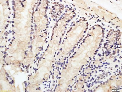
Tissue/cell: rat rectum tissue; 4% Paraformaldehyde-fixed and paraffin-embedded;
Antigen retrieval: citrate buffer ( 0.01M, pH 6.0 ), Boiling bathing for 15min; Block endogenous peroxidase by 3% Hydrogen peroxide for 30min; Blocking buffer (normal goat serum,SLC0005) at 37℃ for 20 min;
Incubation: Anti-XRCC3 Polyclonal Antibody, Unconjugated(SL4261R) 1:100, overnight at 4°C, followed by conjugation to the secondary antibody(SP-0023) and DAB(SLC0010) staining
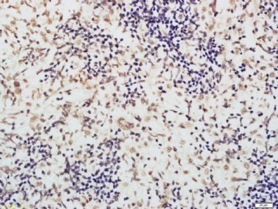
Tissue/cell: rat spleen tissue; 4% Paraformaldehyde-fixed and paraffin-embedded;
Antigen retrieval: citrate buffer ( 0.01M, pH 6.0 ), Boiling bathing for 15min; Block endogenous peroxidase by 3% Hydrogen peroxide for 30min; Blocking buffer (normal goat serum,SLC0005) at 37℃ for 20 min;
Incubation: Anti-XRCC3 Polyclonal Antibody, Unconjugated(SL4261R) 1:100, overnight at 4°C, followed by conjugation to the secondary antibody(SP-0023) and DAB(SLC0010) staining
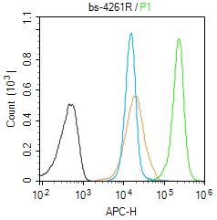
Blank control (Black line):Molt4 (Black).
Primary Antibody (green line): Rabbit Anti-XRCC3 antibody (SL4261R)
Dilution: 1μg /10^6 cells;
Isotype Control Antibody (orange line): Rabbit IgG .
Secondary Antibody (white blue line): Goat anti-rabbit IgG-AF647
Dilution: 1μg /test.
Protocol
The cells were fixed with 4% PFA (10min at room temperature)and then permeabilized with 90% ice-cold methanol for 20 min at room temperature. The cells were then incubated in 5%BSA to block non-specific protein-protein interactions for 30 min at room temperature .Cells stained with Primary Antibody for 30 min at room temperature. The secondary antibody used for 40 min at room temperature. Acquisition of 20,000 events was performed.
|
|
|