Calreticulin is a highly conserved chaperone protein which resides primarily in the endoplasmic reticulum, and is involved in a variety of cellular processes, among them, cell adhesion. Additionally, it functions in protein folding quality control and calcium homeostasis. Calreticulin is also found in the nucleus, suggesting that it may have a role in transcription regulation. Systemic lupus erythematosus is associated with increased autoantibody titers against calreticulin. Recurrent mutations in calreticulin have been linked to various neoplasms, including the myeloproliferative type.[provided by RefSeq, May 2020]
Subunit:
Monomer. Component of an EIF2 complex at least composed of CELF1/CUGBP1, CALR, CALR3, EIF2S1, EIF2S2, HSP90B1 and HSPA5. Interacts with PDIA3/ERp57 (By similarity). Interacts with NR3C1 and TRIM21. Interacts with GABARAP.
Subcellular Location:
Endoplasmic reticulum lumen. Cytoplasm, cytosol. Secreted, extracellular space, extracellular matrix. Cell surface.
Similarity:
Belongs to the calreticulin family.
SWISS:
P27797
Gene ID:
811
Database links:
Entrez Gene: 41166 Fruit fly (Drosophila melanogaster)
Entrez Gene: 811 Human
Entrez Gene: 12317 Mouse
Entrez Gene: 100009050 Rabbit
Entrez Gene: 64202 Rat
Omim: 109091 Human
SwissProt: P29413 Fruit fly (Drosophila melanogaster)
SwissProt: P27797 Human
SwissProt: P14211 Mouse
SwissProt: P15253 Rabbit
SwissProt: P18418 Rat
Unigene: 2457 Fruit fly (Drosophila melanogaster)
Unigene: 515162 Human
Unigene: 1971 Mouse
Unigene: 467043 Mouse
Unigene: 974 Rat
| Picture |
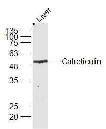
Sample:
Liver (Mouse) Lysate at 40 ug
Primary: Anti-Calreticulin (SL5913R) at 1/1000 dilution
Secondary: IRDye800CW Goat Anti-Rabbit IgG at 1/20000 dilution
Predicted band size: 44 kD
Observed band size: 49 kD
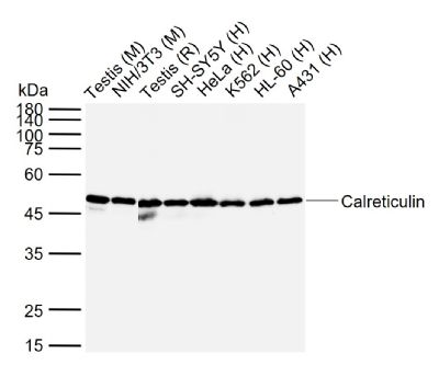
Sample:
Lane 1: Mouse Testis tissue lysates
Lane 2: Mouse NIH/3T3 cell lysates
Lane 3: Rat Testis tissue lysates
Lane 4: Human SH-SY5Y cell lysates
Lane 5: Human HeLa cell lysates
Lane 6: Human K562 cell lysates
Lane 7: Human HL-60 cell lysates
Lane 8: Human A431 cell lysates
Primary: Anti-Calreticulin (SL5913R) at 1/1000 dilution
Secondary: IRDye800CW Goat Anti-Rabbit IgG at 1/20000 dilution
Predicted band size: 44 kDa
Observed band size: 48 kDa
Tissue/cell: rat brain tissue;4% Paraformaldehyde-fixed and paraffin-embedded;
Antigen retrieval: citrate buffer ( 0.01M, pH 6.0 ), Boiling bathing for 15min; Blocking buffer (normal goat serum,SLC0005) at 37℃ for 20 min;
Incubation: Anti-Calreticulin Polyclonal Antibody, FITC conjugated (SL5913R-FITC) 1:100, 40 minutes at 37°C.
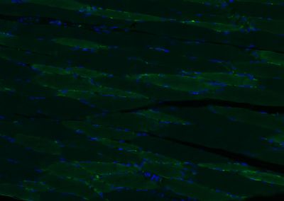
Paraformaldehyde-fixed, paraffin embedded (rat skeletal muscle); Antigen retrieval by boiling in sodium citrate buffer (pH6.0) for 15min; Blocking buffer (normal goat serum) at 37°C for 30min; Antibody incubation with (Calreticulin) Polyclonal Antibody, Unconjugated (SL5913R) at 1:200 overnight at 4°C, followed by a conjugated Goat Anti-Rabbit IgG antibody (SL0295G-AF488) for 90 minutes, and DAPI for nuclei staining.
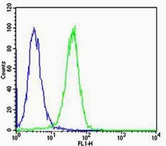
Cell: Hela
Concentration:1:100
Host/Isotype:Rabbit/IgG
Flow cytometric analysis of Rabbit IgG isotype control (Cat#: SL5913R) on Hela(green) compared with control in the absence of primary antibody (blue) followed by Alexa Fluor 488-conjugated goat anti-rabbit IgG(H+L) secondary antibody .
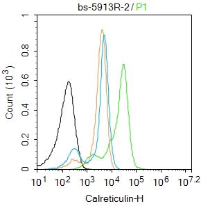
Blank control:K562.
Primary Antibody (green line): Rabbit Anti-Calreticulin antibody (SL5913R)
Dilution: 2μg /10^6 cells;
Isotype Control Antibody (orange line): Rabbit IgG .
Secondary Antibody : Goat anti-rabbit IgG-FITC
Dilution: 0.5μg /test.
Protocol
The cells were fixed with 4% PFA (10min at room temperature)and then permeabilized with 0.1% PBST for 20 min at room temperature. The cells were then incubated in 5%BSA to block non-specific protein-protein interactions for 30 min at room temperature .Cells stained with Primary Antibody for 30 min at room temperature. The secondary antibody used for 40 min at room temperature. Acquisition of 20,000 events was performed.
|
|
|