Home > Product > Antibody > Rabbit Anti-COG1 antibody
Ldlbc; CDG2Gv Component of oligomeric golgi complex 1; Conserved oligomeric Golgi complex protein 1; Low density lipoprotein receptor defect B complementing; COG1_HUMAN.
Cat:
SL6647R
Species Reactivity:
Human,Rat,
Immunogen:
KLH conjugated synthetic peptide derived from human COG1:501-600/980
Format:
Liquid
Storage instructions:
Shipped at 4℃. Store at -20 °C for one year. Avoid repeated freeze/thaw cycles.
Concentration:
1mg/ml
Clonality:
Polyclonal
Isotype:
IgG
Applications:
WB=1:500-2000ELISA=1:5000-10000IHC-P=1:100-500IHC-F=1:100-500IF=1:100-500(Paraffin sections need to do antigen repair)not yet tested in other applications.optimal dilutions/concentrations should be determined by the end user.
Host:
Rabbit
Product Overview:
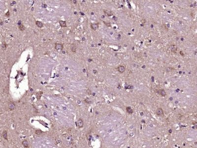
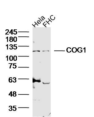
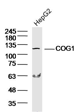
Sample: Hela Cell (Human) Lysate at 40 ugFHC Cell (Human) Lysate at 40 ugPrimary: Anti-COG1 (SL6647R) at 1/300 dilutionSecondary: IRDye800CW Goat Anti-Rabbit IgG at 1/20000 dilutionPredicted band size: 109 kDObserved band size: 109 kDSample: HepG2 Cell (Human) Lysate at 40 ugPrimary: Anti-COG1 (SL6647R) at 1/300 dilutionSecondary: IRDye800CW Goat Anti-Rabbit IgG at 1/20000 dilutionPredicted band size: 109 kDObserved band size: 109 kD Paraformaldehyde-fixed, paraffin embedded (Rat brain); Antigen retrieval by boiling in sodium citrate buffer (pH6.0) for 15min; Block endogenous peroxidase by 3% hydrogen peroxide for 20 minutes; Blocking buffer (normal goat serum) at 37°C for 30min; Antibody incubation with (COG1) Polyclonal Antibody, Unconjugated (SL6647R) at 1:400 overnight at 4°C, followed by operating according to SP Kit(Rabbit) (sp-0023) instructionsand DAB staining.
More
Unit:
Price: $
Product PDFs
Datasheet:


There are eight COG proteins (COG1-8) which form a Golgi-localized complex (COG) required for normal Golgi morphology and function. It is thought that COG1 is required for steps in the normal medial and trans Golgi-associated processing of glycoconjugates and plays a role in the organization of the Golgi-localized complex.

Function:
Required for normal Golgi function (By similarity).

Subunit:
Component of the conserved oligomeric Golgi complex which is composed of eight different subunits and is required for normal Golgi morphology and localization.

Subcellular Location:
Golgi apparatus membrane; Peripheral membrane protein; Cytoplasmic side.

DISEASE:
Defects in COG1 are the cause of congenital disorder of glycosylation type 2G (CDG2G) [MIM:611209]; also known as CDG-II caused by COG1 deficiency. CDGs are a family of severe inherited diseases caused by a defect in glycoprotein biosynthesis. They are characterized by under-glycosylated serum glycoproteins. These multisystem disorders present with a wide variety of clinical features, such as disorders of the nervous system development, psychomotor retardation, dysmorphic features, hypotonia, coagulation disorders and immunodeficiency. The broad spectrum of features reflects the critical role of N-glycoproteins during embryonic development, differentiation, and maintenance of cell functions. Clinical features of CDG2G include failure to thrive, generalized hypotonia, growth retardation and mild psychomotor retardation. CDG2G is biochemically characterized by a defect in O-glycosylation as well as N-glycosylation.

Similarity:
Belongs to the COG1 family.

SWISS:
Q8WTW3

Gene ID:
9382

Database links:

Entrez Gene: 9382 Human

Entrez Gene: 16834 Mouse

Omim: 606973 Human

SwissProt: Q8WTW3 Human

SwissProt: Q9Z160 Mouse

Unigene: 103555 Human

Unigene: 261620 Mouse


Picture

Sample:
Hela Cell (Human) Lysate at 40 ug
FHC Cell (Human) Lysate at 40 ug
Primary: Anti-COG1 (SL6647R) at 1/300 dilution
Secondary: IRDye800CW Goat Anti-Rabbit IgG at 1/20000 dilution
Predicted band size: 109 kD
Observed band size: 109 kD
Sample: HepG2 Cell (Human) Lysate at 40 ug
Primary: Anti-COG1 (SL6647R) at 1/300 dilution
Secondary: IRDye800CW Goat Anti-Rabbit IgG at 1/20000 dilution
Predicted band size: 109 kD
Observed band size: 109 kD
Paraformaldehyde-fixed, paraffin embedded (Rat brain); Antigen retrieval by boiling in sodium citrate buffer (pH6.0) for 15min; Block endogenous peroxidase by 3% hydrogen peroxide for 20 minutes; Blocking buffer (normal goat serum) at 37°C for 30min; Antibody incubation with (COG1) Polyclonal Antibody, Unconjugated (SL6647R) at 1:400 overnight at 4°C, followed by operating according to SP Kit(Rabbit) (sp-0023) instructionsand DAB staining.
Product Feedback Wall
Message :
Your Email :
Copyright © 2007-2018 Sunlong Medical All Rights Reserved.