Binding of FREASLC3 and FREASLC4 to their cognate sites results in bending of the DNA at an angle of 80-90 degrees.
Involvement in disease;
Defects in FOXC1 are the cause of Axenfeld-Rieger syndrome type 3 (RIEG3); also known as Axenfeld-Rieger syndrome (ARS) or Axenfeld syndrome or Axenfeld anomaly. It is characterized by posterior corneal embryotoxon, prominent Schwalbe line and iris adhesion to the Schwalbe line. Other features may be hypertelorism (wide spacing of the eyes), hypoplasia of the malar bones, congenital absence of some teeth and mental retardation. When associated with tooth anomalies, the disorder is known as Rieger syndrome. Glaucoma is a progressive blinding condition that occurs in approximately half of patients with Axenfeld-Rieger malformations.
Function:
Binding of FREASLC3 and FREASLC4 to their cognate sites results in bending of the DNA at an angle of 80-90 degrees.
Subunit:
Monomer.
Subcellular Location:
Nucleus.
Tissue Specificity:
Expressed in all tissues and cell lines examined.
DISEASE:
Defects in FOXC1 are the cause of iridogoniodysgenesis anomaly (IGDA) [MIM:601631]. IGDA is an autosomal dominant phenotype characterized by iris hypoplasia, goniodysgenesis, and juvenile glaucoma.
[DISEASE] Defects in FOXC1 are a cause of Peters anomaly (PAN) [MIM:604229]. Peters anomaly consists of a central corneal leukoma, absence of the posterior corneal stroma and Descemet membrane, and a variable degree of iris and lenticular attachments to the central aspect of the posterior cornea.
Similarity:
Contains 1 fork-head DNA-binding domain.
SWISS:
Q12948
Gene ID:
2296
Database links:
Entrez Gene: 2296 Human
Entrez Gene: 17300 Mouse
GenBank: NP_001444 Human
Omim: 601090 Human
SwissProt: Q12948 Human
SwissProt: Q61572 Mouse
SwissProt: Q32NP8 Xenopus laevis
Unigene: 348883 Human
Unigene: 12949 Mouse
| Picture |
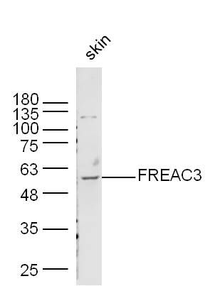
Sample: Skin (Mouse) Lysate at 40 ug
Primary: Anti-FREAC3 (SL6642R) at 1/300 dilution
Secondary: IRDye800CW Goat Anti-Rabbit IgG at 1/20000 dilution
Predicted band size: 57 kD
Observed band size: 57 kD
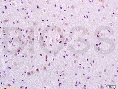
Tissue/cell: rat brain tissue; 4% Paraformaldehyde-fixed and paraffin-embedded;
Antigen retrieval: citrate buffer ( 0.01M, pH 6.0 ), Boiling bathing for 15min; Block endogenous peroxidase by 3% Hydrogen peroxide for 30min; Blocking buffer (normal goat serum,SLC0005) at 37℃ for 20 min;
Incubation: Anti-FOXC1/FREAC3 Polyclonal Antibody, Unconjugated(SL6642R) 1:200, overnight at 4°C, followed by conjugation to the secondary antibody(SP-0023) and DAB(SLC0010) staining
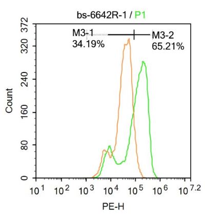
Blank control:A431.
Primary Antibody (green line): Rabbit Anti-FREAC3 antibody (SL6642R)
Dilution: 1μg /10^6 cells;
Isotype Control Antibody (orange line): Rabbit IgG .
Secondary Antibody : Goat anti-rabbit IgG-AF647
Dilution: 1μg /test.
Protocol
The cells were fixed with 4% PFA (10min at room temperature)and then permeabilized with 90% ice-cold methanol for 20 min at-20℃. The cells were then incubated in 5%BSA to block non-specific protein-protein interactions for 30 min at at room temperature .Cells stained with Primary Antibody for 30 min at room temperature. The secondary antibody used for 40 min at room temperature. Acquisition of 20,000 events was performed.
|
|
|