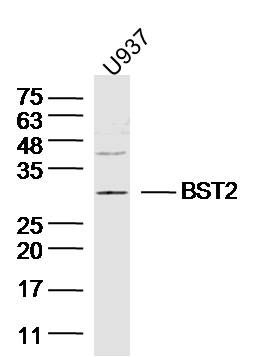
Lane 1: (Human/cell line)U937 lysates probed with BST2 Polyclonal Antibody, Unconjugated (Catalog #SL9850R) at 1:300 overnight at 4°C. Followed by a conjugated secondary antibody (Secondary Catalog #926-32211) at 1:10000 for 60 min at 37°C.
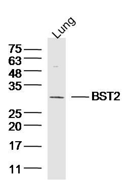
Lane 1: (mouse/tissue)Lung lysates probed with BST2 Polyclonal Antibody, Unconjugated (Catalog #SL9850R) at 1:300 overnight at 4°C. Followed by a conjugated secondary antibody (Secondary Catalog #926-32211) at 1:10000 for 60 min at 37°C.
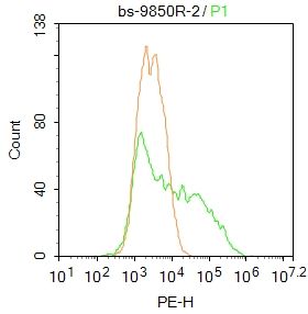
Blank control: A431.
Primary Antibody (green line): Rabbit Anti-BST2 antibody (SL9850R)
Dilution: 2μg /10^6 cells;
Isotype Control Antibody (orange line): Rabbit IgG .
Secondary Antibody : Goat anti-rabbit IgG-PE
Dilution: 1μg /test.
Protocol
The cells were fixed with 4% PFA (10min at room temperature)and then permeabilized with PBST for 20 min at room temperature. The cells were then incubated in 5%BSA to block non-specific protein-protein interactions for 30 min at at room temperature .Cells stained with Primary Antibody for 30 min at room temperature. The secondary antibody used for 40 min at room temperature. Acquisition of 20,000 events was performed.
|