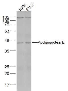
Specific References (5) | SL4892R has been referenced in 5 publications.
[IF=2.942] Xue Gang. et al. Identification of key genes of papillary thyroid carcinoma by integrated bioinformatics analysis. Bioscience Rep. 2020 Aug;40(8):BSR20201555 WB ; Human.
[IF=3.517] Chen YC et al. Indole Compound NC009-1 Augments APOE and TRKA in Alzheimer's Disease Cell and Mouse Models for Neuroprotection and Cognitive Improvement. J Alzheimers Dis. 2019;67(2):737-756. WB ; Human.
[IF=3.412] Chiu CY et al. Fish Oil Supplementation Alleviates the Altered Lipid Homeostasis in Blood, Liver, and Adipose Tissues in High-Fat Diet-Fed Rats.J Agric Food Chem. 2018 Apr 25;66(16):4118-4128. WB ; Rat.
[IF=1.96] Liu, Zan, et al. "Secretomes are a potential source of molecular targets for cancer therapies and indicate that APOE is a candidate biomarker for lung adenocarcinoma metastasis." Molecular Biology Reports (2014): 1-17. WB ; Human.
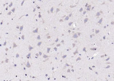
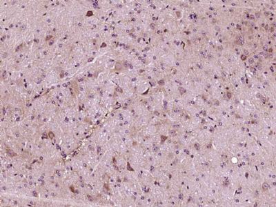
[IF=1.27] Wanwei Jiang. et al. The effect of sevoflurane on the spatial recall ability and expression of apolipoprotein E and β amyloid in the hippocampus in rats. Cell Mol Biol. 2020 Oct;66(7):35-43 IHC ; Rat.