[IF=3.388] Liu H et al. Circular RNA has_circ_000644 inhibits oxLDL-induced vascular endothelial cell proliferation and angiogenesis. Cell Signal. 2020 Jun;70:109595. WB ; human.
[IF=5.572] Liu H et al. Anti-tubulin agent vinorelbine inhibits metastasis of cancer cells by regulating epithelial-mesenchymal transition. Eur J Med Chem. 2020 Aug 15;200:112332. WB ; Human.
[IF=2.073] Jin Dai. et al. NK6 Homeobox 2 Regulated Gastrokin-2 Suppresses Gastric Cancer Cell Proliferation and Invasion via Akt Signaling Pathway. Cell Biochem Biophys. 2021 Mar;79(1):123-131 WB ; Human.
[IF=1.841] Qionghua Hu. et al. Long Non-Coding RNA CASC2 Overexpression Ameliorates Sepsis-Associated Acute Kidney Injury by Regulating MiR-545-3p/PPARA axis. J Surg Res. 2021 Sep;265:223 WB ; Human.
[IF=4.101] Haixiang Ding. et al. Ailanthone suppresses the activity of human colorectal cancer cells through the STAT3 signaling pathway. Int J Mol Med. 2022 Feb;49(2):1-11 WB ; Human.
[IF=10.435] Shi, Yesi. et al. Biomimetic nanoparticles blocking autophagy for enhanced chemotherapy and metastasis inhibition via reversing focal adhesion disassembly. J Nanobiotechnol. 2021 Dec;19(1):1-17 WB ; Human.
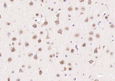
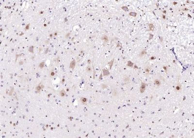
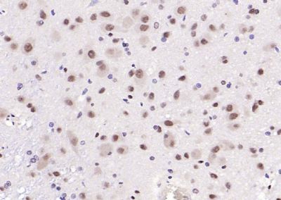
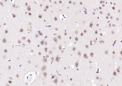
[IF=4.192] Chen D et al. Sodium Propionate Attenuates the Lipopolysaccharide-Induced Epithelial–Mesenchymal Transition via the PI3K/Akt/mTOR Signaling Pathway. J Agric Food Chem. 2020 Jun 17;68(24):6554-6563. IHC ; Mouse.
[IF=10.679] Pan J et al. lncRNA JPX/miR-33a-5p/Twist1 axis regulates tumorigenesis and metastasis of lung cancer by activating Wnt/β-catenin signaling. Mol Cancer. 2020 Jan 15;19(1):9. WB ; Human.
[IF=3.06] Yan Y et al. Inhibition of TGF-β Signaling in Gliomas by the Flavonoid Diosmetin Isolated from Dracocephalum peregrinum L. Molecules. 2020 Jan 2;25(1). pii: E192. WB ; Human.
[IF=5.714] Li D et al. Oxygenated Polycyclic aromatic hydrocarbons (Oxy-PAHs) facilitate lung cancer metastasis by epigenetically regulating the epithelial-to-mesenchymal transition (EMT). Environ Pollut. 2019 Sep 17;255(Pt 2):113261. WB ; Human.
[IF=3.775] Xue Y et al. Chlorogenic acid attenuates cadmium-induced intestinal injury in Sprague–Dawley rats. Food Chem Toxicol. 2019 Aug 4;133:110751. WB ; Rat.
[IF=2.705] Zhou S et al. Helicobacter pylori infection promotes epithelial-to-mesenchymal transition of gastric cells by upregulating LAPTM4B. Biochem Biophys Res Commun. 2019 Jun 30;514(3):893-900. IHSLCP,ICF&WB ; Human.
[IF=3.249] Lin X et al. TFF3 Contributes to Epithelial-Mesenchymal Transition (EMT) in Papillary Thyroid Carcinoma Cells via the MAPK/ERK Signaling Pathway.(2018)J Cancer;Oct 31;9(23):4430-4439. ICC ; Human.
[IF=2.092] Guo F et al. miR-375-3p/YWHAZ/β-catenin axis regulates migration, invasion, EMT in gastric cancer cells.(2018) Clin Exp Pharmacol Physiol. Oct 24. WB ;
[IF=4.357] Fei et al. The number of polyploid giant cancer cells and epithelial-mesenchymal transition-related proteins are associated with invasion and metastasis in human breast cancer. (2015) J.Exp.Clin.Cancer.Res. 34:158 WB ; Human.
[IF=1.28] Jinquan Cai Roles of transcriptional factor Snail and adhesion factor E-cadherin in clear cell renal cell carcinoma. (2013) Exp.Ther.Me. 6:1489-1493 IHSLCP ; Human.
[IF=2.81] Hu et al. Hydroxysafflor Yellow A Ameliorates Renal Fibrosis by Suppressing TGF-β1-Induced Epithelial-to-Mesenchymal Transition. (2016) PLoS.On. 11:e0153409 WB ; Mouse.
[IF=6.1] Yun, Yang, et al. "Sulfate Aerosols Promote Lung Cancer Metastasis by Epigenetically Regulating the Epithelial-to-Mesenchymal Transition (EMT)." Environmental Science & Technology (2017). WB ; Human.
[IF=7.43] Wang, Jing, et al. "Phosphorylation-dependent regulation of ALDH1A1 by Aurora kinase A: insights on their synergistic relationship in pancreatic cancer." BMC biology 15.1 (2017): 10. WB ; Human.
[IF=4.069] Geng X et al. Role of ZIP8 in regulating cell morphology and NF-κB/Snail2 signaling.Metallomics. 2018 Jul 18;10(7):953-964. ICF ; Mouse.
[IF=4.36] Yuhua Li. et al. Bolbostemma paniculatum (Maxim.) Franquet extract suppresses the development of colorectal cancer through downregulation of PI3K/Akt pathway. J Ethnopharmacol. 2022 Apr;287:114937 WB ; Human.
[IF=5.211] Jin, Meiyuan. et al. MicroRNA-3935 promotes human trophoblast cell epithelial-mesenchymal transition through tumor necrosis factor receptor-associated factor 6/regulator of G protein signaling 2 axis. Reprod Biol Endocrin. 2021 Dec;19(1):1-15 WB ; human.
[IF=5.682] Xiaolan You. et al. Fibroblastic galectin-1-fostered invasion and metastasis are mediated by TGF-β1-induced epithelial-mesenchymal transition in gastric cancer. Aging-Us. 2021 Jul 31; 13(14): 18464–18481 WB,IHC ; Mouse.
[IF=4.101] Yu Guo. et al. RepSox effectively promotes the induced differentiation of sheep fibroblasts into adipocytes via the inhibition of the TGF‑β1/Smad pathway. Int J Mol Med. 2021 Aug;48(2):1-13 WB ;
[IF=2.349] Shunichiro Hanai. et al. Hypoxia-induced thyroid hormone receptor expression regulates cell-cycle progression in renal tubule epithelial cells. 2021 Jun 08 IHC ; Human.
[IF=4.147] Francesca Salamanna. et al. Development and characterization of a novel human 3D model of bone metastasis from breast carcinoma in vitro cultured. Bone. 2021 Feb;143:115773 IHC ; Human.
[IF=2.942] Shi M et al. MicroRNA-27a targets Sfrp1 to induce renal fibrosis in diabetic nephropathy by activating Wnt/β-Catenin signalling. Biosci Rep. 2020 Jun 26;40(6):BSR20192794. WB&ICF ; Rat.
[IF=3.04] Ma W et al. A vanillin derivative suppresses the growth of HT29 cells through the Wnt/β-catenin signaling pathway. Eur J Pharmacol. 2019 Apr 15;849:43-49. WB ; Human.
[IF=2.656] Fang et al. Upregulation of long noncoding RNA CCAT1-L promotes epithelial-mesenchymal transition in gastric adenocarcinoma. (2018) Onco.Targets.Ther. 11:5647-5655 WB ; Human.
[IF=1.829] Zhong Y et al. Isolation of primitive mouse extraembryonic endoderm (pXEN) stem cell lines.Stem Cell Res. 2018 Jul;30:100-112. ICF ; Mouse.
[IF=4.43] Wang et al. The Aurora-A-Twist1 axis promotes highly aggressive phenotypes in pancreatic carcinoma. (2017) J.Cell.Sci. 130:1078-1093 WB ; Human.
[IF=4.12] Wang et al. Kukoamine A inhibits human glioblastoma cell growth and migration through apoptosis induction and epithelial-mesenchymal transition attenuation. (2016) Sci.Rep. 6:36543 WB ; Human.
[IF=2.98] Deng et al. PEG10 plays a crucial role in human lung cancer proliferation, progression, prognosis and metastasis. (2014) Oncol.Rep. 32:2159-67 WB ; Human.
[IF=1.238] Liu and Xiao Notch1 signaling induces epithelial-mesenchymal transition in lens epithelium cells during hypoxia. (2017) BMC.Ophthalmo. 17:135 WB ; Human.
[IF=8.332] Short et al. Influenza virus damages the alveolar barrier by disrupting epithelial cell tight junctions. (2016) Eur.Respir.. 47:954-66 IF(ICC) ; Human.
[IF=3.13] Zhang, Wen-feng, et al. "Angelica polysaccharides inhibit the growth and promote the apoptosis of U251 glioma cells in vitro and in vivo." Phytomedicine (2017). WB ; Human.
[IF=3.23] Borin, Thaiz F., et al. "HET0016 decreases lung metastasis from breast cancer in immune-competent mouse model." PLoS One 12.6 (2017). WB, IHSLCP ; Mouse.
[IF=2.25] Neelam, Sudha, Morgan M. Brooks, and Patrick R. Cammarata. "Lenticular cytoprotection, part 2: Link between glycogen synthase kinase-3β, epithelial to mesenchymal transition, and mitochondrial depolarization." (2015) Molecular Vision. Human.
[IF=3.73] Chen, Cheng-Hsien, et al. "MicroRNA-328 Inhibits Renal Tubular Cell Epithelial-to-Mesenchymal Transition by Targeting the CD44 in Pressure-Induced Renal Fibrosis." PloS one 9.6 (2014): e91962. WB ; Rat.