Home > Product > Antibody > Rabbit Anti-MCT1 antibody
monocarboxylate transporter 1; Malignant T cell amplified sequence 1; MCT 1; MCT1; MCTS 1; MCTS1; Oncogene MCT 1; Oncogene MCT1; SLC16A1; FLJ36745; HHF7; MCT; MGC44475; monocarboxylate transporter; Monocarboxylate transporter isoform 1; Monocarboxylic aci
Cat:
SL10249R
Species Reactivity:
Human,Mouse,Rat,(predicted: Dog,Pig,Cow,Horse,Rabbit,Guinea Pig,)
Immunogen:
KLH conjugated synthetic peptide derived from human MCT1:251-350/500<Extracellular>
Format:
Liquid
Storage instructions:
Shipped at 4℃. Store at -20 °C for one year. Avoid repeated freeze/thaw cycles.
Concentration:
1mg/ml
Clonality:
Polyclonal
Isotype:
IgG
Applications:
WB=1:500-2000ELISA=1:5000-10000IHC-P=1:100-500IHC-F=1:100-500Flow-Cyt=1μg/TestIF=1:100-500(Paraffin sections need to do antigen repair)not yet tested in other applications.optimal dilutions/concentrations should be determined by the end user.
Host:
Rabbit
Product Overview:
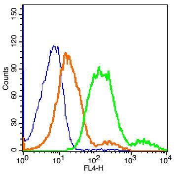
Sample:HepG2 (Human)cell Lysate at 40 ugPrimary: Anti-MCT1(SL10249R)at 1/300 dilutionSecondary: IRDye800CW Goat Anti-RabbitIgG at 1/20000 dilutionPredicted band size: 55kDObserved band size: 55kDSample: Lane 1: Liver (Mouse) Lysate at 40 ugLane 2: Stomach (Mouse) Lysate at 40 ugLane 3: Heart (Mouse) Lysate at 40 ugLane 4: Liver (Rat) Lysate at 40 ugLane 5: Stomach (Rat) Lysate at 40 ugLane 6: Heart (Rat) Lysate at 40 ugPrimary: Anti-MCT1 (SL10249R) at 1/1000 dilutionSecondary: IRDye800CW Goat Anti-Rabbit IgG at 1/20000 dilutionPredicted band size: 48 kDObserved band size: 48 kDParaformaldehyde-fixed, paraffin embedded (rat brain); Antigen retrieval by boiling in sodium citrate buffer (pH6.0) for 15min; Block endogenous peroxidase by 3% hydrogen peroxide for 20 minutes; Blocking buffer (normal goat serum) at 37°C for 30min; Antibody incubation with (MCT1) Polyclonal Antibody, Unconjugated (SL10249R) at 1:400 overnight at 4°C, followed by operating according to SP Kit(Rabbit) (sp-0023) instructionsand DAB staining.Blank control:K562. Primary Antibody (green line): Rabbit Anti-MCT1 antibody (SL10249R) Dilution: 1μg /10^6 cells; Isotype Control Antibody (orange line): Rabbit IgG . Secondary Antibody : Goat anti-rabbit IgG-FITCDilution: 0.5μg /test. ProtocolThe cells were incubated in 5%BSA to block non-specific protein-protein interactions for 30 min at room temperature .Cells stained with Primary Antibody for 30 min at room temperature. The secondary antibody used for 40 min at room temperature. Acquisition of 20,000 events was performed.Blank control: MCF7 Cells(blue). Primary Antibody: Rabbit Anti-MCT1/AF647 Conjugated antibody (SL10249R-AF647), Dilution: 1μg in 100 μL 1X PBS containing 0.5% BSA; Isotype Control Antibody: Rabbit IgG/FITC(orange) ,used under the same conditions.
More
Unit:
Price: $
Product PDFs
Datasheet:


The protein encoded by this gene is a proton-linked monocarboxylate transporter that catalyzes the movement of many monocarboxylates, such as lactate and pyruvate, across the plasma membrane. Mutations in this gene are associated with erythrocyte lactate transporter defect. Alternatively spliced transcript variants have been found for this gene.[provided by RefSeq, Oct 2009]

Function:
Proton-linked monocarboxylate transporter. Catalyzes the rapid transport across the plasma membrane of many monocarboxylates such as lactate, pyruvate, branched-chain oxo acids derived from leucine, valine and isoleucine, and the ketone bodies acetoacetate, beta-hydroxybutyrate and acetate.

Subcellular Location:
Cell membrane; Multi-pass membrane protein.

Tissue Specificity:
Widely expressed in normal and in cancer cells.

DISEASE:
Defects in SLC16A1 are the cause of symptomatic deficiency in lactate transport (SDLT) [MIM:245340]; also known as erythrocyte lactate transporter defect. Deficiency of lactate transporter may result in an acidic intracellular environment created by muscle activity with consequent degeneration of muscle and release of myoglobin and creatine kinase. This defect might compromise extreme performance in otherwise healthy individuals.
Defects in SLC16A1 are the cause of familial hyperinsulinemic hypoglycemia type 7 (HHF7) [MIM:610021]; also known as exercise-induced hyperinsulinemic hypoglycemia. HHF7 is a dominantly inherited hypoglycemic disorder characterized by inappropriate insulin secretion during anaerobic exercise or on pyruvate load.

Similarity:
Belongs to the major facilitator superfamily. Monocarboxylate porter (TC 2.A.1.13) family.

SWISS:
P53985

Gene ID:
6566

Database links:

Entrez Gene: 6566 Human

Entrez Gene: 20501 Mouse

Omim: 600682 Human

SwissProt: P53985 Human

SwissProt: P53986 Mouse

Unigene: 75231 Human

Unigene: 9086 Mouse


Picture

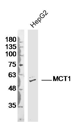
Sample:HepG2 (Human)cell Lysate at 40 ug
Primary: Anti-MCT1(SL10249R)at 1/300 dilution
Secondary: IRDye800CW Goat Anti-RabbitIgG at 1/20000 dilution
Predicted band size: 55kD
Observed band size: 55kD
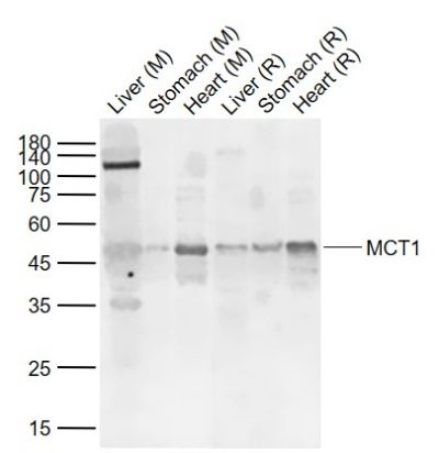
Sample:
Lane 1: Liver (Mouse) Lysate at 40 ug
Lane 2: Stomach (Mouse) Lysate at 40 ug
Lane 3: Heart (Mouse) Lysate at 40 ug
Lane 4: Liver (Rat) Lysate at 40 ug
Lane 5: Stomach (Rat) Lysate at 40 ug
Lane 6: Heart (Rat) Lysate at 40 ug
Primary: Anti-MCT1 (SL10249R) at 1/1000 dilution
Secondary: IRDye800CW Goat Anti-Rabbit IgG at 1/20000 dilution
Predicted band size: 48 kD
Observed band size: 48 kD
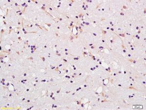
Paraformaldehyde-fixed, paraffin embedded (rat brain); Antigen retrieval by boiling in sodium citrate buffer (pH6.0) for 15min; Block endogenous peroxidase by 3% hydrogen peroxide for 20 minutes; Blocking buffer (normal goat serum) at 37°C for 30min; Antibody incubation with (MCT1) Polyclonal Antibody, Unconjugated (SL10249R) at 1:400 overnight at 4°C, followed by operating according to SP Kit(Rabbit) (sp-0023) instructionsand DAB staining.
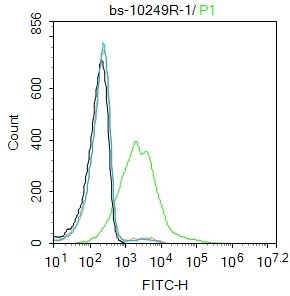
Blank control:K562.
Primary Antibody (green line): Rabbit Anti-MCT1 antibody (SL10249R)
Dilution: 1μg /10^6 cells;
Isotype Control Antibody (orange line): Rabbit IgG .
Secondary Antibody : Goat anti-rabbit IgG-FITC
Dilution: 0.5μg /test.
Protocol
The cells were incubated in 5%BSA to block non-specific protein-protein interactions for 30 min at room temperature .Cells stained with Primary Antibody for 30 min at room temperature. The secondary antibody used for 40 min at room temperature. Acquisition of 20,000 events was performed.
Blank control: MCF7 Cells(blue).
Primary Antibody: Rabbit Anti-MCT1/AF647 Conjugated antibody (SL10249R-AF647), Dilution: 1μg in 100 μL 1X PBS containing 0.5% BSA;
Isotype Control Antibody: Rabbit IgG/FITC(orange) ,used under the same conditions.
Product Feedback Wall
Message :
Your Email :
Copyright © 2007-2018 Sunlong Medical All Rights Reserved.