Home > Product > Antibody > Rabbit Anti-Glucocorticoid Receptor antibody
Glucocorticoid Receptor beta; GCCR; GCR; GCR_HUMAN; glucocorticoid nuclear receptor variant 1; Glucocorticoid receptor beta isoform; GR; GRL; nr3c1; Nuclear receptor subfamily 3 group C member 1.
Cat:
SL13385R
Species Reactivity:
Human,Mouse,Rat,(predicted: Chicken,Dog,Pig,Cow,Horse,Sheep,)
Immunogen:
KLH conjugated synthetic peptide derived from human Glucocorticoid Receptor beta:51-150/777
Format:
Liquid
Storage instructions:
Shipped at 4℃. Store at -20 °C for one year. Avoid repeated freeze/thaw cycles.
Concentration:
1mg/ml
Clonality:
Polyclonal
Isotype:
IgG
Applications:
WB=1:500-2000ELISA=1:5000-10000IHC-P=1:100-500IHC-F=1:100-500Flow-Cyt=1ug/TestICC=1:100-500IF=1:100-500(Paraffin sections need to do antigen repair)not yet tested in other applications.optimal dilutions/concentrations should be determined by the end user.
Host:
Rabbit
Product Overview:
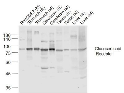
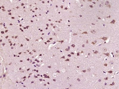
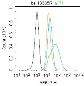
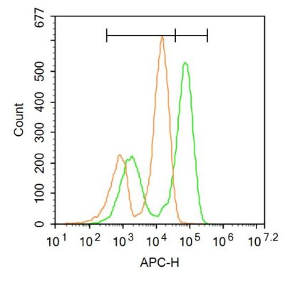
Sample: Lane 1: Raw264.7 (Mouse) Cell Lysate at 30 ugLane 2: Stomach (Rat) Lysate at 40 ugLane 3: Stomach (Mouse) Lysate at 40 ugLane 4: Cerebrum (Rat) Lysate at 40 ugLane 5: Cerebrum (Mouse) Lysate at 40 ugLane 6: Testis (Rat) Lysate at 40 ugLane 7: Testis (Mouse) Lysate at 40 ugLane 8: Liver (Rat) Lysate at 40 ugLane 9: Liver (Mouse) Lysate at 40 ugPrimary: Anti-Glucocorticoid Receptor (SL13385R) at 1/1000 dilutionSecondary: IRDye800CW Goat Anti-Rabbit IgG at 1/20000 dilutionPredicted band size: 90 kDObserved band size: 87 kDParaformaldehyde-fixed, paraffin embedded (mouse brain tissue); Antigen retrieval by boiling in sodium citrate buffer (pH6.0) for 15min; Block endogenous peroxidase by 3% hydrogen peroxide for 20 minutes; Blocking buffer (normal goat serum) at 37°C for 30min; Antibody incubation with (GCR) Polyclonal Antibody, Unconjugated (SL13385R) at 1:400 overnight at 4°C, followed by operating according to SP Kit(Rabbit) (sp-0023) instructionsand DAB staining.Blank control:A549. Primary Antibody (green line): Rabbit Anti-Glucocorticoid Receptor antibody (SL13385R) Dilution: 1μg /10^6 cells; Isotype Control Antibody (orange line): Rabbit IgG . Secondary Antibody : Goat anti-rabbit IgG-AF647Dilution: 1μg /test. ProtocolThe cells were fixed with 4% PFA (10min at room temperature)and then permeabilized with 0.1% PBST for 20 min at room temperature. The cells were then incubated in 5%BSA to block non-specific protein-protein interactions for 30 min at room temperature .Cells stained with Primary Antibody for 30 min at room temperature. The secondary antibody used for 40 min at room temperature. Acquisition of 20,000 events was performed.Blank control:Mouse spleen. Primary Antibody (green line): Rabbit Anti-Glucocorticoid Receptor beta antibody (SL13385R) Dilution: 3μg /10^6 cells; Isotype Control Antibody (orange line): Rabbit IgG . Secondary Antibody: Goat anti-rabbit IgG-AF647Dilution: 3μg /test. ProtocolThe cells were fixed with 4% PFA (10min at room temperature)and then permeabilized with 90% ice-cold methanol for 20 min at-20℃. The cells were then incubated in 5%BSA to block non-specific protein-protein interactions for 30 min at at room temperature .Cells stained with Primary Antibody for 30 min at room temperature. The secondary antibody used for 40 min at room temperature. Acquisition of 20,000 events was performed.
More
Unit:
Price: $
Product PDFs
Datasheet:


Steroid receptors are ligand-dependent, intracellular proteins that stimulate transcription of specific genes by binding to specific DNA sequences following activation by the appropriate hormone. Glucocorticoids are a family of steroids necessary for the regulation of energy metabolism and the immune and inflammatory responses. These compounds exert their effect through their interaction with the glucocoticoid receptor (GR) and that complex's subsequent association with DNA. All normal mammalian tissues examined to date have been shown to contain glucocorticoid receptor.

Function:
Receptor for glucocorticoids (GC). Has a dual mode of action: as a transcription factor that binds to glucocorticoid response elements (GRE) and as a modulator of other transcription factors. Affects inflammatory responses, cellular proliferation and differentiation in target tissues. Could act as a coactivator for STAT5-dependent transcription upon growth hormone (GH) stimulation and could reveal an essential role of hepatic GR in the control of body growth. Involved in chromatin remodeling. Plays a significant role in transactivation. Involved in nuclear translocation.

Subunit:
Heteromultimeric cytoplasmic complex with HSP90, HSP70, and FKBP5 or another immunophilin, or the immunophilin homolog PPP5C. Directly interacts with UNC45A. Upon ligand binding FKBP5 dissociates from the complex and FKBP4 takes its place, thereby linking the complex to dynein and mediating transport to the nucleus, where the complex dissociates (By similarity). Binds to DNA as a homodimer, and as a heterodimer with NR3C2 or the retinoid X receptor. Binds STAT5A and STAT5B homodimers and heterodimers. Interacts with NRIP1, POU2F1, POU2F2 and TRIM28. Interacts with NCOA1, NCOA3, SMARCA4, SMARCC1, SMARCD1, and SMARCE1 (By similarity). Interacts with several coactivator complexes, including the SMARCA4 complex, CREBBP/EP300, TADA2L and p160 coactivators such as NCOA2 and NCOA6. Interaction with BAG1 inhibits transactivation. Interacts with HEXIM1, PELP1 and TGFB1I1.

Subcellular Location:
Cytoplasm. Nucleus. Cytoplasmic in the absence of ligand, nuclear after ligand-binding and Nucleus. Localized largely in the nucleus.

Tissue Specificity:
Widely expressed. In the heart, detected in left and right atria, left and right ventricles, aorta, apex, intraventricular septum, and atrioventricular node as well as whole adult and fetal heart.

Post-translational modifications:
Increased proteasome-mediated degradation in response to glucocorticoids. Phosphorylated in the absence of hormone; becomes hyperphosphorylated in the presence of glucocorticoid. The Ser-203-phosphorylated form is mainly cytoplasmic, and the Ser-211-phosphorylated form is nuclear. Transcriptional activity correlates with the amount of phosphorylation at Ser-211.
Sumoylated; this reduces transcription transactivation.
Ubiquitinated; restricts glucocorticoid-mediated transcriptional signaling.

DISEASE:
Defects in NR3C1 are a cause of glucocorticoid resistance (GCRES) [MIM:27640]; also known as cortisol resistance. It is a hypertensive, hyperandrogenic disorder characterized by increased serum cortisol concentrations. Inheritance is autosomal dominant.

Similarity:
Belongs to the nuclear hormone receptor family. NR3 subfamily.
Contains 1 nuclear receptor DNA-binding domain.

SWISS:
P04150

Gene ID:
2908

Database links:

Entrez Gene: 2908 Human

Entrez Gene: 14815 Mouse

Entrez Gene: 24413 Rat

Omim: 27640 Human

SwissProt: P04150 Human

SwissProt: P06537 Mouse

SwissProt: P06536 Rat

Unigene: 122926 Human

Unigene: 129481 Mouse

Unigene: 90070 Rat


Picture

Sample:
Lane 1: Raw264.7 (Mouse) Cell Lysate at 30 ug
Lane 2: Stomach (Rat) Lysate at 40 ug
Lane 3: Stomach (Mouse) Lysate at 40 ug
Lane 4: Cerebrum (Rat) Lysate at 40 ug
Lane 5: Cerebrum (Mouse) Lysate at 40 ug
Lane 6: Testis (Rat) Lysate at 40 ug
Lane 7: Testis (Mouse) Lysate at 40 ug
Lane 8: Liver (Rat) Lysate at 40 ug
Lane 9: Liver (Mouse) Lysate at 40 ug
Primary: Anti-Glucocorticoid Receptor (SL13385R) at 1/1000 dilution
Secondary: IRDye800CW Goat Anti-Rabbit IgG at 1/20000 dilution
Predicted band size: 90 kD
Observed band size: 87 kD
Paraformaldehyde-fixed, paraffin embedded (mouse brain tissue); Antigen retrieval by boiling in sodium citrate buffer (pH6.0) for 15min; Block endogenous peroxidase by 3% hydrogen peroxide for 20 minutes; Blocking buffer (normal goat serum) at 37°C for 30min; Antibody incubation with (GCR) Polyclonal Antibody, Unconjugated (SL13385R) at 1:400 overnight at 4°C, followed by operating according to SP Kit(Rabbit) (sp-0023) instructionsand DAB staining.
Blank control:A549.
Primary Antibody (green line): Rabbit Anti-Glucocorticoid Receptor antibody (SL13385R)
Dilution: 1μg /10^6 cells;
Isotype Control Antibody (orange line): Rabbit IgG .
Secondary Antibody : Goat anti-rabbit IgG-AF647
Dilution: 1μg /test.
Protocol
The cells were fixed with 4% PFA (10min at room temperature)and then permeabilized with 0.1% PBST for 20 min at room temperature. The cells were then incubated in 5%BSA to block non-specific protein-protein interactions for 30 min at room temperature .Cells stained with Primary Antibody for 30 min at room temperature. The secondary antibody used for 40 min at room temperature. Acquisition of 20,000 events was performed.
Blank control:Mouse spleen.
Primary Antibody (green line): Rabbit Anti-Glucocorticoid Receptor beta antibody (SL13385R)
Dilution: 3μg /10^6 cells;
Isotype Control Antibody (orange line): Rabbit IgG .
Secondary Antibody: Goat anti-rabbit IgG-AF647
Dilution: 3μg /test.
Protocol
The cells were fixed with 4% PFA (10min at room temperature)and then permeabilized with 90% ice-cold methanol for 20 min at-20℃. The cells were then incubated in 5%BSA to block non-specific protein-protein interactions for 30 min at at room temperature .Cells stained with Primary Antibody for 30 min at room temperature. The secondary antibody used for 40 min at room temperature. Acquisition of 20,000 events was performed.
Product Feedback Wall
Message :
Your Email :
Copyright © 2007-2018 Sunlong Medical All Rights Reserved.