Neurofilaments can be defined as the intermediate or 10nm filaments found in specifically in neuronal cells. When visualised using an electron microscope, neurofilaments appear as 10nm diameter fibres of indeterminate length that generally have fine wispy protrusions from their sides. They are particularly abundant in axons of large projection neurons. They probably function to provide structural support for neurons and their synapses and to support the large axon diameters required for rapid conduction of impulses down axons. Neurofilaments are composed of a mixture of subunits, which usually includes the three neurofilament triplet proteins neurofilament light (NFL), neurofilament medium (NFM) and neurofilament heavy (NFH). Neurofilaments may also include smaller amounts of peripherin, alpha internexin, nestin and in some cases vimentin. Antibodies to the various neurofilament subunits are very useful cell type markers since the proteins are among the most abundant of the nervous system, are expressed only in neurons, and are biochemically very stable. Some studies have shown that levels of neurofilament heavy and neurofilament light are elevated in patients with Alzheimer's disease, frontotemporal lobe dementia, and vascular dementia.
Function:
Neurofilaments usually contain three intermediate filament proteins: L, M, and H which are involved in the maintenance of neuronal caliber. NF-H has an important function in mature axons that is not subserved by the two smaller NF proteins.
Post-translational modifications:
There are a number of repeats of the tripeptide K-S-P, NFH is phosphorylated on a number of the serines in this motif. It is thought that phosphorylation of NFH results in the formation of interfilament cross bridges that are important in the maintenance of axonal caliber.
Phosphorylation seems to play a major role in the functioning of the larger neurofilament polypeptides (NF-M and NF-H), the levels of phosphorylation being altered developmentally and coincident with a change in the neurofilament function.
Phosphorylated in the Head and Rod regions by the PKC kinase PKN1, leading to inhibit polymerization.
DISEASE:
Defects in NEFH are a cause of susceptibility to amyotrophic lateral sclerosis (ALS) [MIM:105400]. ALS is a neurodegenerative disorder affecting upper and lower motor neurons, and resulting in fatal paralysis. Sensory abnormalities are absent. Death usually occurs within 2 to 5 years. The etiology is likely to be multifactorial, involving both genetic and environmental factors.
Similarity:
Belongs to the intermediate filament family.
SWISS:
P12036
Gene ID:
4744
Database links:
Entrez Gene: 4744 Human
Entrez Gene: 380684 Mouse
Entrez Gene: 24587 Rat
Omim: 162230 Human
SwissProt: P12036 Human
SwissProt: P19246 Mouse
SwissProt: P16884 Rat
Unigene: 198760 Human
Unigene: 298283 Mouse
Unigene: 108194 Rat
| Picture |
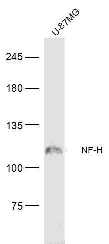
Sample:
U-87MG(Human) Cell Lysate at 30 ug
Primary: Anti-NF-H (SL10136R) at 1/300 dilution
Secondary: IRDye800CW Goat Anti-Rabbit IgG at 1/20000 dilution
Predicted band size: 118 kD
Observed band size: 118 kD
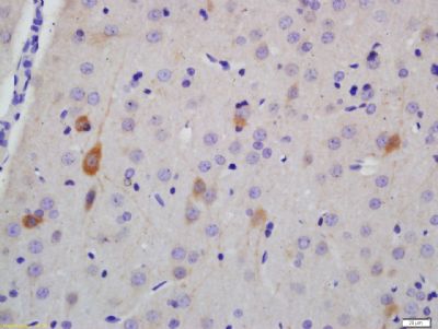
Tissue/cell: rat brain tissue; 4% Paraformaldehyde-fixed and paraffin-embedded;
Antigen retrieval: citrate buffer ( 0.01M, pH 6.0 ), Boiling bathing for 15min; Block endogenous peroxidase by 3% Hydrogen peroxide for 30min; Blocking buffer (normal goat serum,SLC0005) at 37℃ for 20 min;
Incubation: Anti-NF-H Polyclonal Antibody, Unconjugated(SL10136R) 1:200, overnight at 4°C, followed by conjugation to the secondary antibody(SP-0023) and DAB(SLC0010) staining
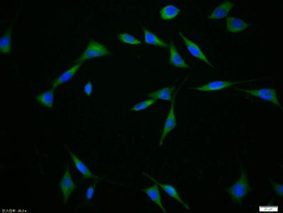
Tissue/cell:SH-SY5Y cell; 4% Paraformaldehyde-fixed; Triton X-100 at room temperature for 20 min; Blocking buffer (normal goat serum,SLC0005) at 37°C for 20 min; Antibody incubation with (NF-H) polyclonal Antibody, Unconjugated (SL10136R) 1:100, 90 minutes at 37°C; followed by a FITC conjugated Goat Anti-Rabbit IgG antibody at 37°C for 90 minutes, DAPI (blue, C02-04002) was used to stain the cell nuclei.
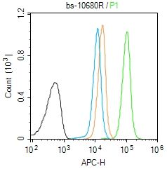
Blank control (Black line): Molt4 (Black).
Primary Antibody (green line):Rabbit Anti-NF-H antibody (SL10136R)
Dilution:1μg /10^6 cells;
Isotype Control Antibody (orange line): Rabbit IgG .
Secondary Antibody (white blue line): Goat anti-rabbit IgG-AF647
Dilution: 1μg /test.
Protocol
The cells were fixed with 4% PFA (10min at room temperature)and then permeabilized with 90% ice-cold methanol for 20 min at room temperature. The cells were then incubated in 5%BSA to block non-specific protein-protein interactions for 30 min at room temperature .Cells stained with Primary Antibody for 30 min at room temperature. The secondary antibody used for 40 min at room temperature. Acquisition of 20,000 events was performed.
|
|
|