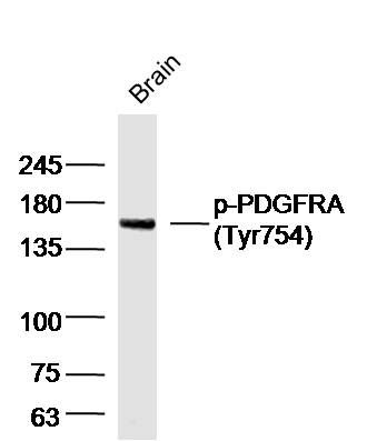
Sample: Brain (Mouse) Lysate at 40 ug
Primary: Anti-Phospho-PDGFRA(Tyr754) (SL20280R) at 1/300 dilution
Secondary: IRDye800CW Goat Anti-Rabbit IgG at 1/20000 dilution
Predicted band size: 117 kD
Observed band size: 150 kD
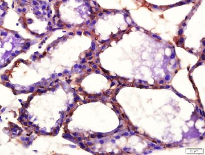
Paraformaldehyde-fixed, paraffin embedded (Rat mammary gland); Antigen retrieval by boiling in sodium citrate buffer (pH6.0) for 15min; Block endogenous peroxidase by 3% hydrogen peroxide for 20 minutes; Blocking buffer (normal goat serum) at 37°C for 30min; Antibody incubation with (Phosphorylated platelet-derived-growth-factor receptor alpha; p-PDGFRA) Polyclonal Antibody, Unconjugated (SL20280R) at 1:400 overnight at 4°C, followed by a conjugated secondary antibody (sp-0023) for 20 minutes and DAB staining.
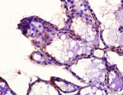
Paraformaldehyde-fixed, paraffin embedded (Rat mammary gland); Antigen retrieval by boiling in sodium citrate buffer (pH6.0) for 15min; Block endogenous peroxidase by 3% hydrogen peroxide for 20 minutes; Blocking buffer (normal goat serum) at 37°C for 30min; Antibody incubation with (Phosphorylated platelet-derived-growth-factor receptor alpha; p-PDGFRA) Polyclonal Antibody, Unconjugated (SL20280R) at 1:400 overnight at 4°C, followed by a conjugated secondary antibody (sp-0023) for 20 minutes and DAB staining.
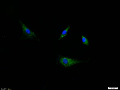
Tissue/cell:A549 cell; 4% Paraformaldehyde-fixed; Triton X-100 at room temperature for 20 min; Blocking buffer (normal goat serum,SLC0005) at 37°C for 20 min; Antibody incubation with (Phospho-PDGFRA (Tyr754)) polyclonal Antibody, Unconjugated (SL20280R) 1:100, 90 minutes at 37°C; followed by a FITC conjugated Goat Anti-Rabbit IgG antibody at 37°C for 90 minutes, DAPI (blue, C02-04002) was used to stain the cell nuclei.
|