Home > Product > Antibody > Rabbit Anti-ITCH/AIP4 antibody
ADMFD; AIF4; AIP4; Atrophin 1 interacting protein 4; Atrophin-1-interacting protein 4; dJ468O1.1; dJ468O1.1 (atrophin 1 interacting protein 4 (AIP4)); dJ468O1.1 atrophin 1 interacting protein 4 AIP4; E3 ubiquitin protein ligase Itchy homolog; E3
Cat:
SL8713R
Species Reactivity:
Human,Mouse,Rat,(predicted: Dog,Pig,Cow,Horse,Rabbit,Sheep,)
Immunogen:
KLH conjugated synthetic peptide derived from human ITCH/AIP4:2-100/903
Format:
Liquid
Storage instructions:
Shipped at 4℃. Store at -20 °C for one year. Avoid repeated freeze/thaw cycles.
Concentration:
1mg/ml
Clonality:
Polyclonal
Isotype:
IgG
Applications:
WB=1:500-2000IHC-P=1:100-500IHC-F=1:100-500ICC=1:100-500IF=1:100-500(Paraffin sections need to do antigen repair)not yet tested in other applications.optimal dilutions/concentrations should be determined by the end user.
Host:
Rabbit
Product Overview:
Sample: MCF-7 Cell (Human) Lysate at 40 ugPrimary: Anti-ITCH/AIP4 (SL8713R) at 1/300 dilutionSecondary: IRDye800CW Goat Anti-Rabbit IgG at 1/20000 dilutionPredicted band size: 103 kDObserved band size: 98 kDParaformaldehyde-fixed, paraffin embedded (Rat brain); Antigen retrieval by boiling in sodium citrate buffer (pH6.0) for 15min; Block endogenous peroxidase by 3% hydrogen peroxide for 20 minutes; Blocking buffer (normal goat serum) at 37°C for 30min; Antibody incubation with (ITCH AIP4) Polyclonal Antibody, Unconjugated (SL8713R) at 1:400 overnight at 4°C, followed by operating according to SP Kit(Rabbit) (sp-0023) instructionsand DAB staining. Paraformaldehyde-fixed, paraffin embedded (Mouse brain); Antigen retrieval by boiling in sodium citrate buffer (pH6.0) for 15min; Block endogenous peroxidase by 3% hydrogen peroxide for 20 minutes; Blocking buffer (normal goat serum) at 37°C for 30min; Antibody incubation with (ITCH) Polyclonal Antibody, Unconjugated (SL8713R) at 1:500 overnight at 4°C, followed by a conjugated secondary (sp-0023) for 20 minutes and DAB staining.
More
Unit:
Price: $
Product PDFs
Datasheet:


Acts as an E3 ubiquitin-protein ligase which accepts ubiquitin from an E2 ubiquitin-conjugating enzyme in the form of a thioester and then directly transfers the ubiquitin to targeted substrates. It catalyzes 'Lys-29'-, 'Lys-48'- and 'Lys-63'-linked ubiquitin conjugation. It is involved in the control of inflammatory signaling pathways. Is an essential component of a ubiquitin-editing protein complex, comprising also TNFAIP3, TAX1BP1 and RNF11, that ensures the transient nature of inflammatory signaling pathways. Promotes the association of the complex after TNF stimulation. Once the complex is formed, TNFAIP3 deubiquitinates 'Lys-63' polyubiquitin chains on RIPK1 and catalyzes the formation of 'Lys-48'-polyubiquitin chains. This leads to RIPK1 proteosomal degradation and consequently termination of the TNF- or LPS-mediated activation of NFKB1. Ubiquitinates RIPK2 by 'Lys-63'-linked conjugation and influences NOD2-dependent signal transduction pathways. Regulates the transcriptional activity of several transcription factors, and probably plays an important role in the regulation of immune response. Ubiquitinates NFE2 by 'Lys-63' linkages and is implicated in the control of the development of hematopoietic lineages. Critical regulator of T helper (TH2) cytokine development through its ability to induce JUNB ubiquitination and degradation (By similarity). Ubiquitinates SNX9. Ubiquitinates CXCR4 and HGS/HRS and regulates sorting of CXCR4 to the degradative pathway. It is involved in the negative regulation of MAVS-dependent cellular antiviral responses. Ubiquitinates MAVS through 'Lys-48'-linked conjugation resulting in MAVS proteosomal degradation. Involved in the regulation of apoptosis and reactive oxygen species levels through the ubiquitination and proteosomal degradation of TXNIP. Mediates the antiapoptotic activity of epidermal growth factor through the ubiquitination and proteosomal degradation of p15 BID. Targets DTX1 for lysosomal degradation and controls NOTCH1 degradation, in the absence of ligand, through 'Lys-29'-linked polyubiquitination.

Function:
Acts as an E3 ubiquitin-protein ligase which accepts ubiquitin from an E2 ubiquitin-conjugating enzyme in the form of a thioester and then directly transfers the ubiquitin to targeted substrates. It catalyzes 'Lys-29'-, 'Lys-48'- and 'Lys-63'-linked ubiquitin conjugation. It is involved in the control of inflammatory signaling pathways. Is an essential component of a ubiquitin-editing protein complex, comprising also TNFAIP3, TAX1BP1 and RNF11, that ensures the transient nature of inflammatory signaling pathways. Promotes the association of the complex after TNF stimulation. Once the complex is formed, TNFAIP3 deubiquitinates 'Lys-63' polyubiquitin chains on RIPK1 and catalyzes the formation of 'Lys-48'-polyubiquitin chains. This leads to RIPK1 proteasomal degradation and consequently termination of the TNF- or LPS-mediated activation of NFKB1. Ubiquitinates RIPK2 by 'Lys-63'-linked conjugation and influences NOD2-dependent signal transduction pathways. Regulates the transcriptional activity of several transcription factors, and probably plays an important role in the regulation of immune response. Ubiquitinates NFE2 by 'Lys-63' linkages and is implicated in the control of the development of hematopoietic lineages. Critical regulator of T-helper (TH2) cytokine development through its ability to induce JUNB ubiquitination and degradation (By similarity). Ubiquitinates SNX9. Ubiquitinates CXCR4 and HGS/HRS and regulates sorting of CXCR4 to the degradative pathway. It is involved in the negative regulation of MAVS-dependent cellular antiviral responses. Ubiquitinates MAVS through 'Lys-48'-linked conjugation resulting in MAVS proteasomal degradation. Involved in the regulation of apoptosis and reactive oxygen species levels through the ubiquitination and proteasomal degradation of TXNIP. Mediates the antiapoptotic activity of epidermal growth factor through the ubiquitination and proteasomal degradation of p15 BID. Targets DTX1 for lysosomal degradation and controls NOTCH1 degradation, in the absence of ligand, through 'Lys-29'-linked polyubiquitination. Ubiquitinates BRAT1 and this ubiquitination is enhanced in the presence of NDFIP1.

Subcellular Location:
Cell membrane. Cytoplasm. Nucleus. Associates with endocytic vesicles. May be recruited to exosomes by NDFIP1.

Tissue Specificity:
Widely expressed.

Post-translational modifications:
On T-cell activation, phosphorylation by the JNK cascade on serine and threonine residues surrounding the PRR domain accelerates the ubiquitination and degradation of JUN and JUNB. The increased ITCH catalytic activity due to phosphorylation by JNK1 may occur due to a conformational change disrupting the interaction between the PRR/WW motifs domain and the HECT domain and, thus exposing the HECT domain (By similarity). Phosphorylation by FYN reduces interaction with JUNB and negatively controls JUN ubiquitination and degradation.
Ubiquitinated; autopolyubiquitination with 'Lys-63' linkages which does not lead to protein degradation.

DISEASE:
Defects in ITCH are the cause of syndromic multisystem autoimmune disease (SMAD) [MIM:613385]. SMAD is characterized by organomegaly, failure to thrive, developmental delay, dysmorphic features and autoimmune inflammatory cell infiltration of the lungs, liver and gut.

Similarity:
Contains 1 C2 domain.
Contains 1 HECT (E6AP-type E3 ubiquitin-protein ligase) domain.
Contains 4 WW domains.

SWISS:
Q96J02

Gene ID:
83737

Database links:

Entrez Gene: 83737 Human

Entrez Gene: 16396 Mouse

Entrez Gene: 311567 Rat

Omim: 601289 Human

SwissProt: Q96J02 Human

SwissProt: Q8C863 Mouse

Unigene: 632272 Human

Unigene: 208286 Mouse

Unigene: 20718 Rat


Picture

Sample:
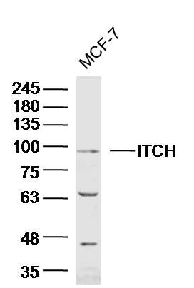
MCF-7 Cell (Human) Lysate at 40 ug
Primary: Anti-ITCH/AIP4 (SL8713R) at 1/300 dilution
Secondary: IRDye800CW Goat Anti-Rabbit IgG at 1/20000 dilution
Predicted band size: 103 kD
Observed band size: 98 kD
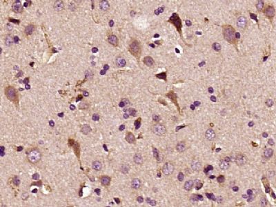
Paraformaldehyde-fixed, paraffin embedded (Rat brain); Antigen retrieval by boiling in sodium citrate buffer (pH6.0) for 15min; Block endogenous peroxidase by 3% hydrogen peroxide for 20 minutes; Blocking buffer (normal goat serum) at 37°C for 30min; Antibody incubation with (ITCH AIP4) Polyclonal Antibody, Unconjugated (SL8713R) at 1:400 overnight at 4°C, followed by operating according to SP Kit(Rabbit) (sp-0023) instructionsand DAB staining.
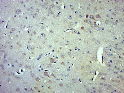
Paraformaldehyde-fixed, paraffin embedded (Mouse brain); Antigen retrieval by boiling in sodium citrate buffer (pH6.0) for 15min; Block endogenous peroxidase by 3% hydrogen peroxide for 20 minutes; Blocking buffer (normal goat serum) at 37°C for 30min; Antibody incubation with (ITCH) Polyclonal Antibody, Unconjugated (SL8713R) at 1:500 overnight at 4°C, followed by a conjugated secondary (sp-0023) for 20 minutes and DAB staining.
Product Feedback Wall
Message :
Your Email :
Copyright © 2007-2018 Sunlong Medical All Rights Reserved.