Home > Product > Antibody > Rabbit Anti-SHISA2 antibody
pProtein shisa 5; Putative NF kappa B activating protein 120; Putative NFkB activating protein 120; Scotin; Shisa homolog 5 (Xenopus laevis).
Cat:
SL24152R
Species Reactivity:
Mouse,
Immunogen:
KLH conjugated synthetic peptide derived from mouse SHISA2 :1-100/235<Extracellular>
Format:
Liquid
Storage instructions:
Shipped at 4℃. Store at -20 °C for one year. Avoid repeated freeze/thaw cycles.
Concentration:
1mg/ml
Clonality:
Polyclonal
Isotype:
IgG
Applications:
WB=1:500-2000IHC-P=1:100-500IHC-F=1:100-500ICC=1:100-500IF=1:100-500(Paraffin sections need to do antigen repair)not yet tested in other applications.optimal dilutions/concentrations should be determined by the end user.
Host:
Rabbit
Product Overview:
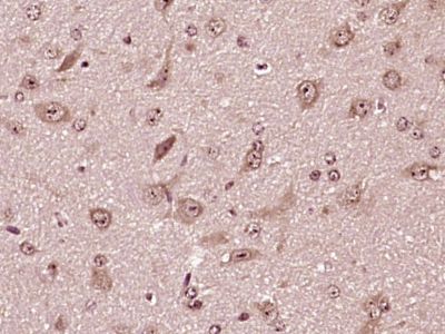
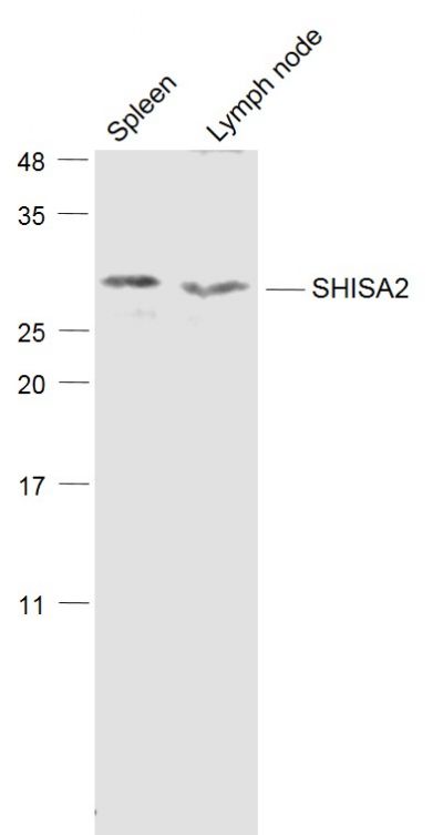
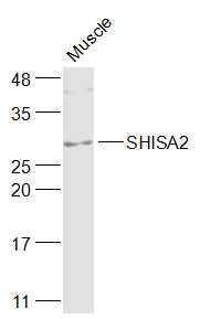
Sample: Spleen (Mouse) Lysate at 40 ugLymph node (Mouse) Lysate at 40 ugPrimary: Anti-SHISA2 (SL24152R) at 1/1000 dilutionSecondary: IRDye800CW Goat Anti-Rabbit IgG at 1/20000 dilutionPredicted band size: 31 kDObserved band size: 31 kDSample: Muscle (Mouse) Lysate at 40 ugPrimary: Anti-SHISA2 (SL24152R) at 1/1000 dilutionSecondary: IRDye800CW Goat Anti-Rabbit IgG at 1/20000 dilutionPredicted band size: 31 kDObserved band size: 31 kDParaformaldehyde-fixed, paraffin embedded (mouse brain tissue); Antigen retrieval by boiling in sodium citrate buffer (pH6.0) for 15min; Block endogenous peroxidase by 3% hydrogen peroxide for 20 minutes; Blocking buffer (normal goat serum) at 37°C for 30min; Antibody incubation with (SHISA2) Polyclonal Antibody, Unconjugated (SL24152R) at 1:400 overnight at 4°C, followed by operating according to SP Kit(Rabbit) (sp-0023) instructionsand DAB staining.
More
Unit:
Price: $
Product PDFs
Datasheet:


This gene encodes a member of the shisa family. The encoded protein is localized to the endoplasmic reticulum, and together with p53 induces apoptosis in a caspase-dependent manner. Alternative splicing results in multiple transcript variants. [provided by RefSeq, Jan 2013]

Function:
Plays an essential role in the maturation of presomitic mesoderm cells by individual attenuation of both FGF and WNT signaling.

Subcellular Location:
Endoplasmic reticulum membrane; Single-pass type I membrane protein. Nucleus membrane.

Similarity:
Belongs to the shisa family.

SWISS:
Q9D7I0

Gene ID:
66940

Database links:

Entrez Gene: 51246 Human

Omim: 607290 Human

SwissProt: Q8N114 Human

Unigene: 414579 Human


Picture

Sample:
Spleen (Mouse) Lysate at 40 ug
Lymph node (Mouse) Lysate at 40 ug
Primary: Anti-SHISA2 (SL24152R) at 1/1000 dilution
Secondary: IRDye800CW Goat Anti-Rabbit IgG at 1/20000 dilution
Predicted band size: 31 kD
Observed band size: 31 kD
Sample:
Muscle (Mouse) Lysate at 40 ug
Primary: Anti-SHISA2 (SL24152R) at 1/1000 dilution
Secondary: IRDye800CW Goat Anti-Rabbit IgG at 1/20000 dilution
Predicted band size: 31 kD
Observed band size: 31 kD
Paraformaldehyde-fixed, paraffin embedded (mouse brain tissue); Antigen retrieval by boiling in sodium citrate buffer (pH6.0) for 15min; Block endogenous peroxidase by 3% hydrogen peroxide for 20 minutes; Blocking buffer (normal goat serum) at 37°C for 30min; Antibody incubation with (SHISA2) Polyclonal Antibody, Unconjugated (SL24152R) at 1:400 overnight at 4°C, followed by operating according to SP Kit(Rabbit) (sp-0023) instructionsand DAB staining.
Product Feedback Wall
Message :
Your Email :
Copyright © 2007-2018 Sunlong Medical All Rights Reserved.