| Picture |
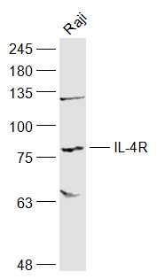
Sample:
Raji(Human) Cell Lysate at 30 ug
Primary: Anti-IL-4R (SL23578R) at 1/1000 dilution
Secondary: IRDye800CW Goat Anti-Rabbit IgG at 1/20000 dilution
Predicted band size: 88 kD
Observed band size: 88 kD
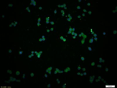
Tissue/cell: 293T cell; 4% Paraformaldehyde-fixed; Triton X-100 at room temperature for 20 min; Blocking buffer (normal goat serum, SLC0005) at 37°C for 20 min; Antibody incubation with (IL-4R) Polyclonal Antibody, Unconjugated (SL23578R) 1:100, 90 minutes at 37°C; followed by a conjugated Goat Anti-Rabbit IgG antibody (SL0295G-FITC) at 37°C for 90 minutes, DAPI (5ug/ml, blue, SLC0033) was used to stain the cell nuclei.
Tissue/cell: 293T cell; 4% Paraformaldehyde-fixed; Triton X-100 at room temperature for 20 min; Blocking buffer (normal goat serum, SLC0005) at 37°C for 20 min; Antibody incubation with (IL-4R) Polyclonal Antibody, Unconjugated (SL23578R) 1:100, 90 minutes at 37°C; followed by a conjugated Goat Anti-Rabbit IgG antibody (SL0295G-FITC) at 37°C for 90 minutes, DAPI (5ug/ml, blue, SLC0033) was used to stain the cell nuclei.
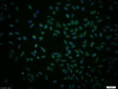
Tissue/cell: A431 cell; 4% Paraformaldehyde-fixed; Triton X-100 at room temperature for 20 min; Blocking buffer (normal goat serum, SLC0005) at 37°C for 20 min; Antibody incubation with (IL-4R) Polyclonal Antibody, Unconjugated (SL23578R) 1:100, 90 minutes at 37°C; followed by a conjugated Goat Anti-Rabbit IgG antibody (SL0295G-FITC) at 37°C for 90 minutes, DAPI (5ug/ml, blue, SLC0033) was used to stain the cell nuclei.
|