Home > Product > Antibody > Rabbit Anti-Cathepsin D antibody
Cathepsin D heavy chain; CatD; CathepsinD; Cathepsin-D; CLN10; CPSD; CTSD; Lysosomal aspartyl peptidase; MGC2311; CATD_HUMAN.
Cat:
SL23781R
Species Reactivity:
Mouse,(predicted: Rat,)
Immunogen:
KLH conjugated synthetic peptide derived from mouse Cathepsin D heavy chain:121-220/410
Format:
Liquid
Storage instructions:
Shipped at 4℃. Store at -20 °C for one year. Avoid repeated freeze/thaw cycles.
Concentration:
1mg/ml
Clonality:
Polyclonal
Isotype:
IgG
Applications:
WB=1:500-2000ELISA=1:5000-10000IHC-P=1:100-500IHC-F=1:100-500ICC=1:100-500IF=1:100-500(Paraffin sections need to do antigen repair)not yet tested in other applications.optimal dilutions/concentrations should be determined by the end user.
Host:
Rabbit
Product Overview:
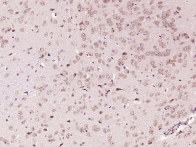
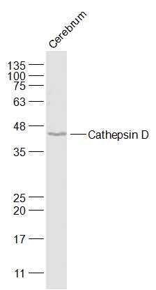
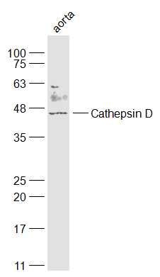
Sample: Cerebrum (Mouse) Lysate at 40 ugPrimary: Anti-Cathepsin D (SL23781R) at 1/1000 dilutionSecondary: IRDye800CW Goat Anti-Rabbit IgG at 1/20000 dilutionPredicted band size: 45 kDObserved band size: 45 kDSample: Aorta(Mouse) Lysate at 40 ugPrimary: Anti-Cathepsin D (SL23781R) at 1/1000 dilutionSecondary: IRDye800CW Goat Anti-Rabbit IgG at 1/20000 dilutionPredicted band size: 45 kDObserved band size: 45 kD Paraformaldehyde-fixed, paraffin embedded (Mouse brain); Antigen retrieval by boiling in sodium citrate buffer (pH6.0) for 15min; Block endogenous peroxidase by 3% hydrogen peroxide for 20 minutes; Blocking buffer (normal goat serum) at 37°C for 30min; Antibody incubation with (Cathepsin D) Polyclonal Antibody, Unconjugated (SL23781R) at 1:400 overnight at 4°C, followed by operating according to SP Kit(Rabbit) (sp-0023) instructionsand DAB staining.
More
Unit:
Price: $
Product PDFs
Datasheet:


Cathepsin D is a normal lysosomal protease that is expressed in all cells. It is an aspartyl protease with a pH optimum in the range of 3-5, and contains two N-linked oligosaccharides. Cathepsin D is synthesized as an inactive 52 kDa pro enzyme. Activation involves the proteolytic removal of the 43 amino acid profragment and an internal cleavage to generate the two-chain form made up of 34 and 14 kDa subunits. Cathepsin D contains the mannose-6-phosphate lysosomal localization signal that targets the enzyme to the lysosomal compartment where it functions in the normal degradation of proteins. In certain tumor cells, Cathepsin D is abnormally processed and is secreted in its 52 kDa precursor form. Numerous clinical studies as well as in vitro evidence suggest that cathepsin D plays an important role in malignant transformation and may be a useful prognostic indicator for breast cancer and possibly Alzheimer's disease.

Function:
Acid protease active in intracellular protein breakdown. Involved in the pathogenesis of several diseases such as breast cancer and possibly Alzheimer disease.

Subcellular Location:
Lysosome. Melanosome. Identified by mass spectrometry in melanosome fractions from stage I to stage IV.

Tissue Specificity:
Expressed in the aorta extrcellular space (at protein level).

Post-translational modifications:
N- and O-glycosylated.

DISEASE:
Defects in CTSD are the cause of neuronal ceroid lipofuscinosis type 10 (CLN10); also known as neuronal ceroid lipofuscinosis due to cathepsin D deficiency. A form of neuronal ceroid lipofuscinosis with onset at birth or early childhood. Neuronal ceroid lipofuscinoses are progressive neurodegenerative, lysosomal storage diseases characterized by intracellular accumulation of autofluorescent liposomal material, and clinically by seizures, dementia, visual loss, and/or cerebral atrophy.

Similarity:
Belongs to the peptidase A1 family.

SWISS:
P18242

Gene ID:
13033

Database links:

Entrez Gene: 1509 Human

Entrez Gene: 13033 Mouse

Omim: 116168 Human

SwissProt: P07339 Human

SwissProt: P18242 Mouse

Unigene: 654447 Human

Unigene: 231395 Mouse


Picture

Sample:
Cerebrum (Mouse) Lysate at 40 ug
Primary: Anti-Cathepsin D (SL23781R) at 1/1000 dilution
Secondary: IRDye800CW Goat Anti-Rabbit IgG at 1/20000 dilution
Predicted band size: 45 kD
Observed band size: 45 kD
Sample:
Aorta(Mouse) Lysate at 40 ug
Primary: Anti-Cathepsin D (SL23781R) at 1/1000 dilution
Secondary: IRDye800CW Goat Anti-Rabbit IgG at 1/20000 dilution
Predicted band size: 45 kD
Observed band size: 45 kD
Paraformaldehyde-fixed, paraffin embedded (Mouse brain); Antigen retrieval by boiling in sodium citrate buffer (pH6.0) for 15min; Block endogenous peroxidase by 3% hydrogen peroxide for 20 minutes; Blocking buffer (normal goat serum) at 37°C for 30min; Antibody incubation with (Cathepsin D) Polyclonal Antibody, Unconjugated (SL23781R) at 1:400 overnight at 4°C, followed by operating according to SP Kit(Rabbit) (sp-0023) instructionsand DAB staining.
Product Feedback Wall
Message :
Your Email :
Copyright © 2007-2018 Sunlong Medical All Rights Reserved.