Home > Product > Antibody > Rabbit Anti-ERK5 antibody
Big MAP kinase 1; BMK 1; BMK 1 kinase; BMK-1; BMK1; BMK1 Kinase; EC 2.7.11.24; ERK 4; ERK 5; ERK-5; ERK4; Extracellular Signal Regulated Kinase 5; Extracellular signal-regulated kinase 5; MAP kinase 7; MAPK 7; MAPK7; Mitogen Activated Protein Kinase 7; Mi
Cat:
SLM52069R
Species Reactivity:
Human,Mouse,Rat,
Immunogen:
Recombinant human ERK5 protein, around C-terminal 200aa
Format:
Liquid
Storage instructions:
Shipped at 4℃. Store at -20 °C for one year. Avoid repeated freeze/thaw cycles.
Concentration:
1mg/ml
Clonality:
Monoclonal
Isotype:
IgG
Applications:
WB=1:500-2000ELISA=1:5000-10000IHC-P=1:100-500Flow-Cyt=2ug/TestICC=1:50-100IF=1:100-500(Paraffin sections need to do antigen repair)not yet tested in other applications.optimal dilutions/concentrations should be determined by the end user.
Host:
Rabbit
Product Overview:
Blocking buffer: 5% NFDM/TBSTPrimary Ab dilution: 1:2000Primary Ab incubation condition: 2 hours atroom temperatureSecondary Ab: Goat Anti-Rabbit IgG H&L(HRP)Lysate: 1: MCF-7, 2: 293T, 3: NIH/3T3, 4: PSLC12Protein loading quantity: 20 μgExposure time: 60 sPredicted MW: 88 kDaObserved MW: 115 kDaSample: Testis (Mouse) Lysate at 40 ugHela(Human) Cell Lysate at 30 ugPrimary: Anti- ERK5 (SLM52069R) at 1/1000 dilutionSecondary: IRDye800CW Goat Anti-Rabbit IgG at 1/20000 dilutionPredicted band size: 49 kDObserved band size: 110 kDParaformaldehyde-fixed, paraffin embedded (rat testis); Antigen retrieval by boiling in sodium citrate buffer (pH6.0) for 15min; Block endogenous peroxidase by 3% hydrogen peroxide for 20 minutes; Blocking buffer (normal goat serum) at 37°C for 30min; Antibody incubation with (ERK5) Polyclonal Antibody, Unconjugated (SLM52069R) at 1:200 overnight at 4°C, followed by operating according to SP Kit(Rabbit) (sp-0023) instructionsand DAB staining.Blank control:MCF7. Primary Antibody (green line): Rabbit Anti-ERK5 antibody (SLM52069R) Dilution: 2μg /10^6 cells; Isotype Control Antibody (orange line): Rabbit IgG . Secondary Antibody : Goat anti-rabbit IgG-AF488Dilution: 1μg /test. ProtocolThe cells were fixed with 4% PFA (10min at room temperature)and then permeabilized with 90% ice-cold methanol for 20 min at-20℃. The cells were then incubated in 5%BSA to block non-specific protein-protein interactions for 30 min at room temperature .Cells stained with Primary Antibody for 30 min at room temperature. The secondary antibody used for 40 min at room temperature. Acquisition of 20,000 events was performed.
More
Unit:
Price: $
Product PDFs
Datasheet:


The protein encoded by this gene is a member of the MAP kinase family. MAP kinases act as an integration point for multiple biochemical signals, and are involved in a wide variety of cellular processes such as proliferation, differentiation, transcription regulation and development. This kinase is specifically activated by mitogen-activated protein kinase kinase 5 (MAP2K5/MEK5). It is involved in the downstream signaling processes of various receptor molecules including receptor type kinases, and G protein-coupled receptors. In response to extracelluar signals, this kinase translocates to cell nucleus, where it regulates gene expression by phosphorylating, and activating different transcription factors. Four alternatively spliced transcript variants of this gene encoding two distinct isoforms have been reported. [provided by RefSeq, Jul 2008]

Function:
Plays a role in various cellular processes such as proliferation, differentiation and cell survival. The upstream activator of MAPK7 is the MAPK kinase MAP2K5. Upon activation, it translocates to the nucleus and phosphorylates various downstream targets including MEF2C. EGF activates MAPK7 through a Ras-independent and MAP2K5-dependent pathway. May have a role in muscle cell differentiation. May be important for endothelial function and maintenance of blood vessel integrity. MAP2K5 and MAPK7 interact specifically with one another and not with MEK1/ERK1 or MEK2/ERK2 pathways. Phosphorylates SGK1 at Ser-78 and this is required for growth factor-induced cell cycle progression.

Subunit:
Interacts with MAP2K5. Forms oligomers. Interacts with MEF2A, MEF2C and MEF2D; the interaction phosphorylates the MEF2s and enhances transcriptional activity of MEF2A, MEF2C but not MEF2D. Interacts with SGK1.

Subcellular Location:
Cytoplasm. Nucleus. Note=Translocates to the nucleus upon activation.

Tissue Specificity:
Expressed in many adult tissues. Abundant in heart, placenta, lung, kidney and skeletal muscle. Not detectable in liver.

Post-translational modifications:
Dually phosphorylated on Thr-219 and Tyr-221, which activates the enzyme. Autophosphorylated in vitro on threonine and tyrosine residues when the SLCterminal part of the kinase, which could have a regulatory role, is absent.

DISEASE:
Note=MAPK is overexpressed in highly metastatic breast cancer cell lines and its expression is preferentially associated with basal-like and metastatic phenotypes of breast tumor samples.

Similarity:
Belongs to the protein kinase superfamily. CMGC Ser/Thr protein kinase family. MAP kinase subfamily.
Contains 1 protein kinase domain.

SWISS:
Q13164

Gene ID:
5598

Database links:

Entrez Gene: 5598 Human

Entrez Gene: 23939 Mouse

Entrez Gene: 114509 Rat

Omim: 602521 Human

SwissProt: Q13164 Human

SwissProt: Q9WVS8 Mouse

SwissProt: P0C865 Rat

Unigene: 150136 Human

Unigene: 38172 Mouse

Unigene: 144629 Rat


Picture

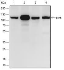
Blocking buffer: 5% NFDM/TBST
Primary Ab dilution: 1:2000
Primary Ab incubation condition: 2 hours at
room temperature
Secondary Ab: Goat Anti-Rabbit IgG H&L
(HRP)
Lysate: 1: MCF-7, 2: 293T, 3: NIH/3T3, 4: PSLC
12
Protein loading quantity: 20 μg
Exposure time: 60 s
Predicted MW: 88 kDa
Observed MW: 115 kDa
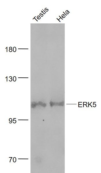
Sample:
Testis (Mouse) Lysate at 40 ug
Hela(Human) Cell Lysate at 30 ug
Primary: Anti- ERK5 (SLM52069R) at 1/1000 dilution
Secondary: IRDye800CW Goat Anti-Rabbit IgG at 1/20000 dilution
Predicted band size: 49 kD
Observed band size: 110 kD
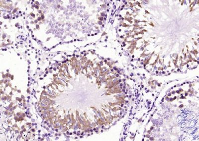
Paraformaldehyde-fixed, paraffin embedded (rat testis); Antigen retrieval by boiling in sodium citrate buffer (pH6.0) for 15min; Block endogenous peroxidase by 3% hydrogen peroxide for 20 minutes; Blocking buffer (normal goat serum) at 37°C for 30min; Antibody incubation with (ERK5) Polyclonal Antibody, Unconjugated (SLM52069R) at 1:200 overnight at 4°C, followed by operating according to SP Kit(Rabbit) (sp-0023) instructionsand DAB staining.
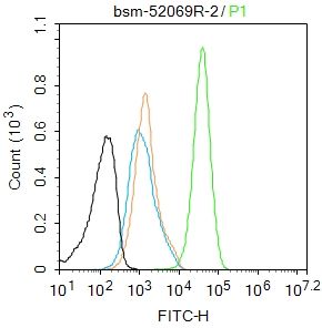
Blank control:MCF7.
Primary Antibody (green line): Rabbit Anti-ERK5 antibody (SLM52069R)
Dilution: 2μg /10^6 cells;
Isotype Control Antibody (orange line): Rabbit IgG .
Secondary Antibody : Goat anti-rabbit IgG-AF488
Dilution: 1μg /test.
Protocol
The cells were fixed with 4% PFA (10min at room temperature)and then permeabilized with 90% ice-cold methanol for 20 min at-20℃. The cells were then incubated in 5%BSA to block non-specific protein-protein interactions for 30 min at room temperature .Cells stained with Primary Antibody for 30 min at room temperature. The secondary antibody used for 40 min at room temperature. Acquisition of 20,000 events was performed.
Product Feedback Wall
Message :
Your Email :
Copyright © 2007-2018 Sunlong Medical All Rights Reserved.