The product of this gene is a component of the nuclear factor of activated T cells DNA-binding transcription complex. This complex consists of at least two components: a preexisting cytosolic component that translocates to the nucleus upon T cell receptor (TCR) stimulation, and an inducible nuclear component. Proteins belonging to this family of transcription factors play a central role in inducible gene transcription during immune response. The product of this gene is an inducible nuclear component. It functions as a major molecular target for the immunosuppressive drugs such as cyclosporin A. Multiple alternatively spliced transcript variants encoding distinct isoforms have been identified for this gene. Different isoforms of this protein may regulate inducible expression of different cytokine genes. [provided by RefSeq, Jul 2013]
Function:
Plays a role in the inducible expression of cytokine genes in T-cells, especially in the induction of the IL-2 or IL-4 gene transcription. Also controls gene expression in embryonic cardiac cells. Could regulate not only the activation and proliferation but also the differentiation and programmed death of T-lymphocytes as well as lymphoid and non-lymphoid cells.Required for osteoclastogenesis and regulates many genes important for osteoclast differentiation and function.
Subunit:
Member of the multicomponent NFATC transcription complex that consists of at least two components, a pre-existing cytoplasmic component NFATC2 and an inducible nuclear component NFATC1. Other members such as NFATC4, NFATC3 or members of the activating protein-1 family, MAF, GATA4 and Cbp/p300 can also bind the complex. NFATC proteins bind to DNA as monomers.
Subcellular Location:
Cytoplasm. Nucleus. Cytoplasmic for the phosphorylated form and nuclear after activation that is controlled by calcineurin-mediated dephosphorylation. Rapid nuclear exit of NFATC is thought to be one mechanism by which cells distinguish between sustained and transient calcium signals. The subcellular localization of NFATC plays a key role in the regulation of gene transcription.
Tissue Specificity:
Expressed in thymus, peripheral leukocytes as T-cells and spleen. Isoforms A are preferentially expressed in effector T-cells (thymus and peripheral leukocytes) whereas isoforms B and isoforms C are preferentially expressed in naive T-cells (spleen). Isoforms B are expressed in naive T-cells after first antigen exposure and isoforms A are expressed in effector T-cells after second antigen exposure.
Post-translational modifications:
Phosphorylated by NFATSLCkinase; dephosphorylated by calcineurin.
Similarity:
Contains 1 RHD (Rel-like) domain.
SWISS:
O95644
Gene ID:
4772
Database links:
Entrez Gene: 4772 Human
Entrez Gene: 3618 Mouse
Entrez Gene: 100361818 Rat
Omim: 600489 Human
SwissProt: O95644 Human
SwissProt: O88942 Mouse
Unigene: 534074 Human
Unigene: 701518 Human
Unigene: 329560 Mouse
Unigene: 229098 Rat
| Picture |
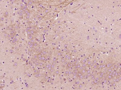
Paraformaldehyde-fixed, paraffin embedded (Rat brain); Antigen retrieval by boiling in sodium citrate buffer (pH6.0) for 15min; Block endogenous peroxidase by 3% hydrogen peroxide for 20 minutes; Blocking buffer (normal goat serum) at 37°C for 30min; Antibody incubation with (NFAT2) Polyclonal Antibody, Unconjugated (SL1417R) at 1:400 overnight at 4°C, followed by operating according to SP Kit(Rabbit) (sp-0023) instructionsand DAB staining.
Blank control: Jurkat.
Primary Antibody (green line): Rabbit Anti-NFAT2 antibody (SL1417R)
Dilution: 1ug/Test;
Secondary Antibody : Goat anti-rabbit IgG-FITC
Dilution: 0.5ug/Test.
Protocol
The cells were fixed with 4% PFA (10min at room temperature)and then permeabilized with 90% ice-cold methanol for 20 min at -20℃.The cells were then incubated in 5%BSA to block non-specific protein-protein interactions for 30 min at room temperature .Cells stained with Primary Antibody for 30 min at room temperature. The secondary antibody used for 40 min at room temperature. Acquisition of 20,000 events was performed.
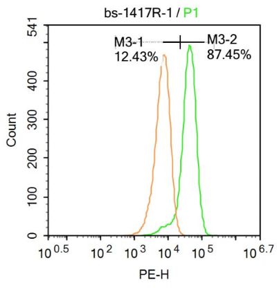
Blank control:Molt-4.
Primary Antibody (green line): Rabbit Anti-NFAT2 antibody (SL1417R)
Dilution: 1μg /10^6 cells;
Isotype Control Antibody (orange line): Rabbit IgG .
Secondary Antibody : Goat anti-rabbit IgG-AF647
Dilution: 1μg /test.
Protocol
The cells were fixed with 4% PFA (10min at room temperature)and then permeabilized with 90% ice-cold methanol for 20 min at-20℃. The cells were then incubated in 5%BSA to block non-specific protein-protein interactions for 30 min at at room temperature .Cells stained with Primary Antibody for 30 min at room temperature. The secondary antibody used for 40 min at room temperature. Acquisition of 20,000 events was performed.
|
|
|