Home > Product > Antibody > Rabbit Anti-VPAC2R antibody
VIP receptor II; VIP receptor2; VIP receptor-2; PACAP type-3; VIP type-2 receptor; Vipr2; Vip2; VPAC2; VPAC2R; Vasoactive intestinal polypeptide receptor 2; VIP-R-2; Pituitary adenylate cyclase-activating polypeptide type III receptor; PACAP type III rece
Cat:
SL0197R
Species Reactivity:
Human,(predicted: Mouse,Rat,Rabbit,)
Immunogen:
KLH conjugated synthetic peptide derived from rat VIP receptor II:81-170/437<Extracellular>
Format:
Liquid
Storage instructions:
Shipped at 4℃. Store at -20 °C for one year. Avoid repeated freeze/thaw cycles.
Concentration:
1mg/ml
Clonality:
Polyclonal
Isotype:
IgG
Applications:
WB=1:500-2000ELISA=1:5000-10000IHC-P=1:100-500IHC-F=1:100-500Flow-Cyt=2ug/TestIF=1:100-500(Paraffin sections need to do antigen repair)not yet tested in other applications.optimal dilutions/concentrations should be determined by the end user.
Host:
Rabbit
Product Overview:
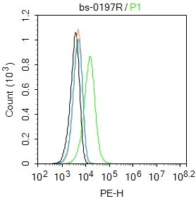
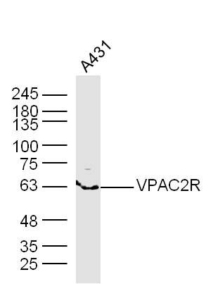
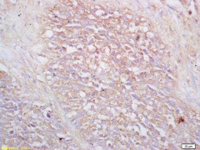
Sample: A431 Cell Lysate at 40 ugPrimary: Anti- VPAC2R (SL0197R) at 1/300 dilutionSecondary: IRDye800CW Goat Anti-Rabbit IgG at 1/20000 dilutionPredicted band size: 48 kDObserved band size: 63 kDTissue/cell: human lung carcinoma; 4% Paraformaldehyde-fixed and paraffin-embedded; Antigen retrieval: citrate buffer ( 0.01M, pH 6.0 ), Boiling bathing for 15min; Block endogenous peroxidase by 3% Hydrogen peroxide for 30min; Blocking buffer (normal goat serum,SLC0005) at 37℃ for 20 min; Incubation: Anti-VIP receptor 2/VPAC2 Polyclonal Antibody, Unconjugated(SL0197R) 1:200, overnight at 4°C, followed by conjugation to the secondary antibody(SP-0023) and DAB(SLC0010) stainingBlank control:Hela. Primary Antibody (green line): Rabbit Anti-VPAC2R antibody (SL0197R) Dilution: 2μg /10^6 cells; Isotype Control Antibody (orange line): Rabbit IgG . Secondary Antibody : Goat anti-rabbit IgG-PEDilution: 2μg /test. ProtocolThe cells were fixed with 4% PFA (10min at room temperature)and then permeabilized with PBST for 20 min at room temperature. The cells were then incubated in 5%BSA to block non-specific protein-protein interactions for 30 min at room temperature .Cells stained with Primary Antibody for 30 min at room temperature. The secondary antibody used for 40 min at room temperature. Acquisition of 20,000 events was performed.
More
Unit:
Price: $
Product PDFs
Datasheet:


Pituitary adenylate cyclase-activating polypeptide (PACAP) is a neuropeptide belonging to the vasoactive intestinal polypeptide/glucagon/ secretin family. It is widely distributed in the body, and a variety of biological actions have been reported. Recent studies have shown that there is a family of PACAP receptors (PACAPRs), and two members of this family have been identified. Mouse PACAPR-3 is a protein of 437 amino acids that has 50% and 51% identity with rat PACAP type I and type II receptors, respectively. It binds to vasoactive intestinal polypeptide as well as PACAP-38 and -27, with a slightly higher affinity for PACAP-38, PACAPR-3 mRNA is expressed at high levels in MIN6, at moderate levels in pancreatic islets and other insulin-secreting cell lines, HIT-T15 and RINm5F, as well as in the lung, brain, stomach, and colon, and at low levels in the heart. PACAPR-3 participates in the regulation of insulin secretion. insulin secretion from MIN6 cells is significantly stimulated by PACAP-38.

Function:
This is a receptor for VIP as well as PACAP-38 and -27, the activity of this receptor is mediated by G proteins which activate adenylyl cyclase. Can be coupled to phospholipase C.

Subcellular Location:
Cell membrane; Multi-pass membrane protein.

Tissue Specificity:
Expressed in CD4+ T-cells, but not in CD8+ T-cells. Expressed in the T-cell lines Jurkat, PEER, MOLT-4, HSB, YT and Tsup-1, but not in the T-cell lines HARRIS and HUT 78.

Similarity:
Belongs to the G-protein coupled receptor 2 family.

SWISS:
P35000

Gene ID:
7434

Database links:

Entrez Gene: 7434 Human

Omim: 601970 Human

SwissProt: P41587 Human

Unigene: 585052 Human


PACAP 受体分为两个亚型,PACAP-2型受体对PACAP是特异性的,与PACAP的亲和力比VIP高100-1000倍,PACAP-II型受体与PACAP和VIP的亲和力相似,对二者无选择性。此抗体即为后者。 Picture

Sample: A431 Cell Lysate at 40 ug
Primary: Anti- VPAC2R (SL0197R) at 1/300 dilution
Secondary: IRDye800CW Goat Anti-Rabbit IgG at 1/20000 dilution
Predicted band size: 48 kD
Observed band size: 63 kD
Tissue/cell: human lung carcinoma; 4% Paraformaldehyde-fixed and paraffin-embedded;
Antigen retrieval: citrate buffer ( 0.01M, pH 6.0 ), Boiling bathing for 15min; Block endogenous peroxidase by 3% Hydrogen peroxide for 30min; Blocking buffer (normal goat serum,SLC0005) at 37℃ for 20 min;
Incubation: Anti-VIP receptor 2/VPAC2 Polyclonal Antibody, Unconjugated(SL0197R) 1:200, overnight at 4°C, followed by conjugation to the secondary antibody(SP-0023) and DAB(SLC0010) staining
Blank control:Hela.
Primary Antibody (green line): Rabbit Anti-VPAC2R antibody (SL0197R)
Dilution: 2μg /10^6 cells;
Isotype Control Antibody (orange line): Rabbit IgG .
Secondary Antibody : Goat anti-rabbit IgG-PE
Dilution: 2μg /test.
Protocol
The cells were fixed with 4% PFA (10min at room temperature)and then permeabilized with PBST for 20 min at room temperature. The cells were then incubated in 5%BSA to block non-specific protein-protein interactions for 30 min at room temperature .Cells stained with Primary Antibody for 30 min at room temperature. The secondary antibody used for 40 min at room temperature. Acquisition of 20,000 events was performed.
Product Feedback Wall
Message :
Your Email :
Copyright © 2007-2018 Sunlong Medical All Rights Reserved.