The placental-derived growth factor (PIGF) is a dimeric glycoprotein showing a high degree of sequence similarity to the vascular endothelial growth factor. Alternative splicing of the PIGF primary transcript gives rise to two forms, named PIGF-1 and PIGF-2, which differ only in the insertion of a highly basic 21-amino acid stretch at the carboxyl end. The presence of the PIGF mRNA in thyroid, placenta, lung, and goiter has indicated the tissues where this factor functions. However, the role of PIGF in vascular development has not yet been clearly established.
Function: Growth factor active in angiogenesis and endothelial cell growth, stimulating their proliferation and migration. It binds to the receptor FLT1/VEGFR-1. Isoform PlGF-2 binds NRP1/neuropilin-1 and NRP2/neuropilin-2 in a heparin-dependent manner.
Subunit: Antiparallel homodimer; disulfide-linked. Also found as heterodimer with VEGFA/VEGF (By similarity).
Subcellular Location: Secreted. The three isoforms are secreted but PlGF-2 appears to remain cell attached unless released by heparin.
Tissue Specificity: While the three isoforms are present in most placental tissues, PlGF-2 is specific to early (8 week) placenta and only PlGF-1 is found in the colon and mammary carcinomas.
Similarity: Belongs to the PDGF/VEGF growth factor family.
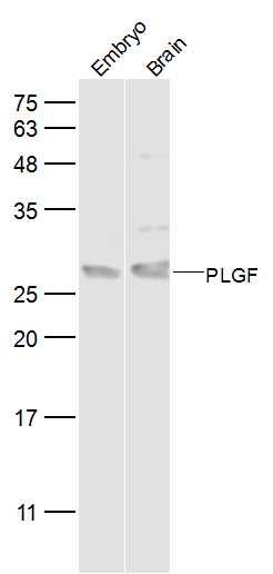
Sample:
Embryo (Mouse) Lysate at 40 ug
Brain (Mouse) Lysate at 40 ug
Primary: Anti-PLGF (SL0234R) at 1/500 dilution
Secondary: IRDye800CW Goat Anti-Rabbit IgG at 1/20000 dilution
Predicted band size: 16 kD
Observed band size: 27 kD
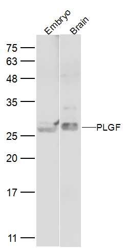
Sample:
Embryo (Mouse) Lysate at 40 ug
Brain (Mouse) Lysate at 40 ug
Primary:Anti-PLGF (SL0234R) at 1/2000 dilution
Secondary: IRDye800CW Goat Anti-Rabbit IgG at 1/20000 dilution
Predicted band size: 16 kD
Observed band size: 27 kD