PHD3 (Egl nine homolog 3; Hypoxia-inducible factor prolyl hydroxylase 3; HIF-prolyl hydroxylase 3; HIF-PH3; HPH-1; Egln3; Prolyl hydroxylase domain-containing protein 3;)Catalyzes the post-translational formation of 4-hydroxyproline in hypoxia-inducible factor (HIF) alpha proteins. Hydroxylates HIF-1 alpha at 'Pro-564', and HIF-2 alpha. Functions as a cellular oxygen sensor and, under normoxic conditions, targets HIF through the hydroxylation for proteasomal degradation via the von Hippel-Lindau ubiquitination complex. May play a role in cell growth regulation in muscle cells and in apoptosis in neuronal tissue. Promotes cell death through a caspase-dependent mechanism. [Catalytic Activity] An HIF alpha chain L-proline + 2-oxoglutarate+ O(2) = An HIF alpha chain trans-4-hydroxy-L-proline + succinate + CO(2). [Subcellular Location] Cytoplasm. Nucleus. Widely expressed at low levels. Expressed athigher levels in heart (cardiac myocytes, aortic endothelial cells and coronary artery smooth muscle) and placenta.
Function:
Cellular oxygen sensor that catalyzes, under normoxic conditions, the post-translational formation of 4-hydroxyproline in hypoxia-inducible factor (HIF) alpha proteins. Hydroxylates a specific proline found in each of the oxygen-dependent degradation (ODD) domains (N-terminal, NODD, and SLCterminal, CODD) of HIF1A. Also hydroxylates HIF2A. Has a preference for the CODD site for both HIF1A and HIF2A. Hydroxylation on the NODD site by EGLN3 appears to require prior hydroxylation on the CODD site. Hydroxylated HIFs are then targeted for proteasomal degradation via the von Hippel-Lindau ubiquitination complex. Under hypoxic conditions, the hydroxylation reaction is attenuated allowing HIFs to escape degradation resulting in their translocation to the nucleus, heterodimerization with HIF1B, and increased expression of hypoxy-inducible genes. EGLN3 is the most important isozyme in limiting physiological activation of HIFs (particularly HIF2A) in hypoxia. Also hydroxylates PKM in hypoxia, limiting glycolysis. Under normoxia, hydroxylates and regulates the stability of ADRB2. Regulator of cardiomyocyte and neuronal apoptosis. In cardiomyocytes, inhibits the anti-apoptotic effect of BCL2 by disrupting the BAX-BCL2 complex. In neurons, has a NGF-induced proapoptotic effect, probably through regulating CASP3 activity. Also essential for hypoxic regulation of neutrophilic inflammation. Plays a crucial role in DNA damage response (DDR) by hydroxylating TELO2, promoting its interaction with ATR which is required for activation of the ATR/CHK1/p53 pathway. Target proteins are preferencially recognized via a LXXLAP motif.
Subunit:
Interacts with WDR83; the interaction leads to almost complete elimination of HIF-mediated reporter activity. Interacts with BCL2 (via its SH4 domain); the interaction disrupts the BAX-BCL4 complex inhibiting the anti-apoptotic activity of BCL2. Interacts with ADRB2; the interaction hydroxylates ADRB2 facilitating its ubiquitination by the VHL-E3 ligase complex. Interacts with PAX2; the interaction targets PAX2 for destruction. Interacts with PKM; the interaction hydroxylates PKM in hypoxia.
Subcellular Location:
Nucleus. Cytoplasm. Note=Colocalizes with WDR83 in the cytoplasm.
Tissue Specificity:
Widely expressed at low levels. Expressed at higher levels in adult heart (cardiac myocytes, aortic endothelial cells and coronary artery smooth muscle), lung and placenta, and in fetal spleen, heart and skeletal muscle. Also expressed in pancreas. Localized to pancreatic acini and islet cells.
Similarity:
Contains 1 Fe2OG dioxygenase domain.
SWISS:
Q9H6Z9
Gene ID:
112399
Database links:
Entrez Gene: 112399 Human
Entrez Gene: 11487 Mouse
Entrez Gene: 54702 Rat
Omim: 606426 Human
SwissProt: Q9H6Z9 Human
SwissProt: Q91UZ4 Mouse
SwissProt: Q62630 Rat
Unigene: 135507 Human
Unigene: 133037 Mouse
Unigene: 10994 Rat
| Picture |
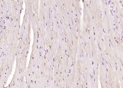
Paraformaldehyde-fixed, paraffin embedded (mouse heart tissue); Antigen retrieval by boiling in sodium citrate buffer (pH6.0) for 15min; Block endogenous peroxidase by 3% hydrogen peroxide for 20 minutes; Blocking buffer (normal goat serum) at 37°C for 30min; Antibody incubation with (PHD3) Polyclonal Antibody, Unconjugated (SL0532R) at 1:400 overnight at 4°C, followed by operating according to SP Kit(Rabbit) (sp-0023) instructionsand DAB staining.
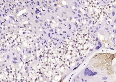
Paraformaldehyde-fixed, paraffin embedded (mouse placenta tissue); Antigen retrieval by boiling in sodium citrate buffer (pH6.0) for 15min; Block endogenous peroxidase by 3% hydrogen peroxide for 20 minutes; Blocking buffer (normal goat serum) at 37°C for 30min; Antibody incubation with (PHD3) Polyclonal Antibody, Unconjugated (SL0532R) at 1:400 overnight at 4°C, followed by operating according to SP Kit(Rabbit) (sp-0023) instructionsand DAB staining.
|
|
|