Home > Product > Antibody > Rabbit Anti-SYAP1 antibody
Synapse associated protein-1; synapse-associated protein 1-like; 2010110O17Rik, AW011796, DKFZp686K221, FLJ14495, FLJ44185, MGC94115, PRO3113, SYAP1; SYAP1_HUMAN; SAP-1.
Cat:
SL0121R
Species Reactivity:
Human,Rat,(predicted: Mouse,Chicken,Dog,Pig,Cow,Horse,)
Immunogen:
KLH conjugated synthetic peptide derived from human SAP-1 :151-230/352
Format:
Liquid
Storage instructions:
Shipped at 4℃. Store at -20 °C for one year. Avoid repeated freeze/thaw cycles.
Concentration:
1mg/ml
Clonality:
Polyclonal
Isotype:
IgG
Applications:
ELISA=1:5000-10000IHC-P=1:100-500IHC-F=1:100-500Flow-Cyt=1μg/TestIF=1:100-500(Paraffin sections need to do antigen repair)not yet tested in other applications.optimal dilutions/concentrations should be determined by the end user.
Host:
Rabbit
Product Overview:
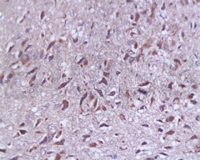
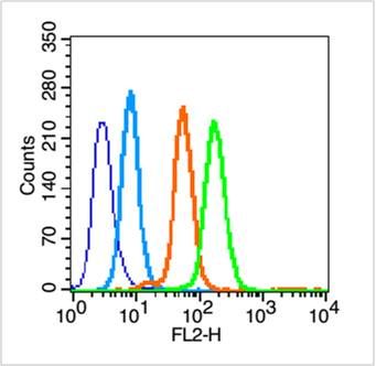
Tissue/cell: rat brain tissue; 4% Paraformaldehyde-fixed and paraffin-embedded; Antigen retrieval: citrate buffer ( 0.01M, pH 6.0 ), Boiling bathing for 15min; Block endogenous peroxidase by 3% Hydrogen peroxide for 30min; Blocking buffer (normal goat serum,SLC0005) at 37℃ for 20 min; Incubation: Anti-SYP p38/Synaptophysin/SAP-1 Polyclonal Antibody, Unconjugated(SL0121R) 1:200, overnight at 4°C, followed by conjugation to the secondary antibody(SP-0023) and DAB(SLC0010) stainingBlank control (blue line): Hep G2 (blue). Primary Antibody (green line): Rabbit Anti-SYAP1 antibody (SL0121R) Dilution: 1μg /10^6 cells; Isotype Control Antibody (orange line): Rabbit IgG . Secondary Antibody (white blue line): Goat anti-rabbit IgG-PEDilution: 1μg /test. ProtocolThe cells were fixed with 70% ethanol (Overnight at 4℃) and then permeabilized with 0.1% PBS-Tween for 20 min at room temperature. Cells stained with Primary Antibody for 30 min at room temperature. The cells were then incubated in 1 X PBS/2%BSA/10% goat serum to block non-specific protein-protein interactions followed by the antibody for 15 min at room temperature. The secondary antibody used for 40 min at room temperature. Acquisition of 20,000 events was performed.
More
Unit:
Price: $
Product PDFs
Datasheet:


SYAP1 (synapse-associated protein 1) is a 352 amino acid protein that is ubiquitously expressed in adult tissues. SYAP1 contains one BSD domain which is a novel domain that is present in basal transcription factors, synapse-associated proteins and several hypothetical proteins. The BSD domain is characterized by three predicted alpha helices and by conserved tryptophan and phenylalanine residues, located at the SLCterminus of the domain. The gene that encodes SYAP1 in humans is located on chromosome X. Chromosome X consists of about 153 million base pairs and nearly 1,000 genes. Color blindness, hemophilia, and Duchenne muscular dystrophy are well known X chromosome-linked conditions which affect males more frequently as males carry a single X chromosome.

Similarity:
Contains 1 BSD domain.

SWISS:
Q96A49

Gene ID:
94056

Database links:

Entrez Gene: 509355 Cow

Entrez Gene: 94056 Human

Entrez Gene: 67043 Mouse

Entrez Gene: 302678 Rat

SwissProt: Q96A49 Human

SwissProt: Q9D5V6 Mouse

Unigene: 729197 Human

Unigene: 44207 Mouse


突触素SAP-1/SYP(Synaptophysin, FRG-4)是分布于突触囊泡膜上含量丰富的糖蛋白之一,与递质释放关系密切,并可影响突触可塑性。突触相关蛋白-1可作为神经元和神经内分泌细胞的可靠标记物用于神经系统形态、功能和发育以及相关疾病的研究。对于SAP-1的功能及其具体作用机制,尚待进一步研究。 Picture

Tissue/cell: rat brain tissue; 4% Paraformaldehyde-fixed and paraffin-embedded;
Antigen retrieval: citrate buffer ( 0.01M, pH 6.0 ), Boiling bathing for 15min; Block endogenous peroxidase by 3% Hydrogen peroxide for 30min; Blocking buffer (normal goat serum,SLC0005) at 37℃ for 20 min;
Incubation: Anti-SYP p38/Synaptophysin/SAP-1 Polyclonal Antibody, Unconjugated(SL0121R) 1:200, overnight at 4°C, followed by conjugation to the secondary antibody(SP-0023) and DAB(SLC0010) staining
Blank control (blue line): Hep G2 (blue).
Primary Antibody (green line): Rabbit Anti-SYAP1 antibody (SL0121R)
Dilution: 1μg /10^6 cells;
Isotype Control Antibody (orange line): Rabbit IgG .
Secondary Antibody (white blue line): Goat anti-rabbit IgG-PE
Dilution: 1μg /test.
Protocol
The cells were fixed with 70% ethanol (Overnight at 4℃) and then permeabilized with 0.1% PBS-Tween for 20 min at room temperature. Cells stained with Primary Antibody for 30 min at room temperature. The cells were then incubated in 1 X PBS/2%BSA/10% goat serum to block non-specific protein-protein interactions followed by the antibody for 15 min at room temperature. The secondary antibody used for 40 min at room temperature. Acquisition of 20,000 events was performed.
Product Feedback Wall
Message :
Your Email :
Copyright © 2007-2018 Sunlong Medical All Rights Reserved.