[IF=6.344] Sun, Lizhong. et al. Fibroblast membrane-camouflaged nanoparticles for inflammation treatment in the early stage. Int J Oral Sci. 2021 Nov;13(1):1-14 FC ; Human.
[IF=3.263] Lina D. Eissa. et al. Inhibition of thioredoxin-interacting protein and inflammasome assembly using verapamil mitigates diabetic retinopathy and pancreatic injury. Eur J Pharmacol. 2021 Mar;:174061 IHC ; Rat.
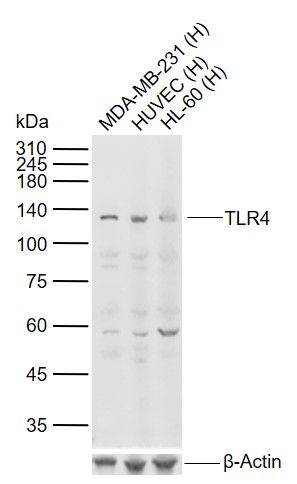
[IF=2.942] Cheng Z et al. Cotinine inhibits TLR4/NF-κB signaling pathway and improves deep vein thrombosis in rats. Biosci Rep. 2020 Jun 26;40(6):BSR20201293. WB ; Rat.
[IF=1.531] Nakayama Y et al. Receptor activator of the NFκB ligand system protects renal function during experimental renal ischemia-reperfusion in mice. Transpl Immunol. 2020 Feb;58:101263. WB ; Mouse.
[IF=4.868] Li Z et al. Methane-Rich Saline Counteracts Cholestasis-Induced Liver Damage via Regulating the TLR4/NF-κB/NLRP3 Inflammasome Pathway. Oxidative Medicine and Cellular Longevity, 2019, 1–13. IHF ; Rat.
[IF=4.868] Wang G et al. Protective Effect of Methane-Rich Saline on Acetic Acid-Induced Ulcerative Colitis via Blockingthe TLR4/NF-κB/MAPK Pathway and Promoting IL-10/JAK1/STAT3-Mediated Anti-inflammatory Response. Oxid Med Cell Longev. 2019 Apr 28;2019:7850324. WB ; Mouse.
[IF=3.47] Li T et al. Milk fat globule membrane supplementation modulates the gut microbiota and attenuates metabolic endotoxemia in high-fat diet-fed mice. Journal of Functional Foods,2018 47, 56–65. WB ; Mouse.
[IF=1.69] Wang et al. Inhibitory effect of Zanthoxylum bungeanum seed oil on ovalbumin?induced lung inflammation in a murine model of asthma. (2016) Mol.Med.Rep. 13:4289-302 WB ; Mouse.
[IF=5.076] Liu L et al. Deficiency of Tenascin-C Alleviates Neuronal Apoptosis and Neuroinflammation After Experimental Subarachnoid Hemorrhage in Mice. Mol Neurobiol. 2018 Nov;55(11):8346-8354. WB ; Mouse.
[IF=0] Arfian et al. Vitamin D Attenuates Kidney Fibrosis via Reducing Fibroblast Expansion, Inflammation, and Epithelial Cell Apoptosis. (2016) Kobe.J.Med.Sci. 62:E38-44 IHSLCP ; Rat.
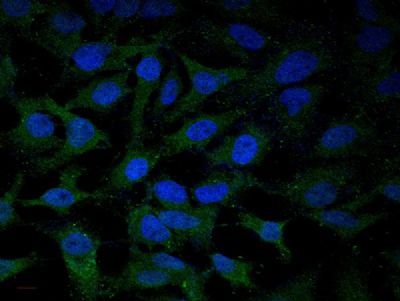
[IF=3.418] Zhao et al. Inhibition of TLR4 Signalling-Induced Inflammation Attenuates Secondary Injury after Diffuse Axonal Injury in Rats. (2016) Mediators.Inflamm. 2016:4706915 IF ; Rat.
[IF=0] Jenvey, Caitlin J., and Judith R. Stabel. "Sudan Black B masks Mycobacterium avium subspecies paratuberculosis immunofluorescent antibody labeling." Journal of Histology & Histopathology 4.1 (2017): 11. IHSLCP ; Bovine.
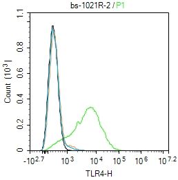
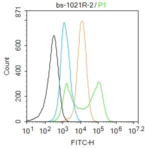
[IF=3.94] Kulhankova, Katarina, et al. "The Superantigen Toxic Shock Syndrome Toxin-1 Alters Human Aortic Endothelial Cell Function." Infection and immunity (2017): IAI-00848. FCM ; Human.
[IF=5.4] Kawakita, Fumihiro, et al. "Effects of Toll-Like Receptor 4 Antagonists Against Cerebral Vasospasm After Experimental Subarachnoid Hemorrhage in Mice." Molecular Neurobiology (2016): 1-10. WB ; Mouse.
[IF=0] ARFIAN, NUR, et al. "Vitamin D Attenuates Kidney Fibrosis via Reducing Fibroblast Expansion, Inflammation, and Epithelial Cell Apoptosis." Kobe J. Med. Sci 62.2 (2016): E38-E44. IHSLCP ; Rat.
[IF=4.52] Namisaki, Tadashi, et al. "Beneficial effects of combined ursodeoxycholic acid and angiotensin-II type 1 receptor blocker on hepatic fibrogenesis in a rat model of nonalcoholic steatohepatitis." Journal of Gastroenterology (2015): 1-11. IHSLCP ; Rat.
[IF=1.92] Wang, Dunjing, et al. "Artesunate Attenuates Lipopolysaccharide-Stimulated Proinflammatory Responses by Suppressing TLR4, MyD88 Expression, and NF-κB Activation in Microglial Cells." Inflammation: 1-8. WB ; Mouse.
[IF=0.1] Xing, Shanshan, et al. "Relationship between toll-like receptor 4 levels in aorta and severity of atherosclerosis." Journal of International Medical Research (2014): 0300060514534645. IHSLCP ; Human.
[IF=1.55] Tang, Lu, et al. "Expression of TRAF6 and pro-inflammatory cytokines through activation of TLR2, TLR4, NOD1, and NOD2 in human periodontal ligament fibroblasts." Archives of Oral Biology 56.10 (2011): 1064-1072. Human.
[IF=3.48] Chen, Xiaoming, et al. ʺSargassum fusiforme polysaccharide activates nuclear factor kappa-B (NF-κB) and induces cytokine production via Toll-like receptors.ʺ Carbohydrate Polymers (2014). other ; Rat.
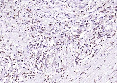
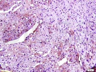
[IF=2.967] Lili Li. et al. Protein overexpression of toll‑like receptor 4 and myeloid differentiation factor 88 in oral squamous cell carcinoma and clinical significance. Oncol Lett. 2021 Nov;22(5):1-11 IHC ; human.
[IF=4.187] Jiajia Wang. et al. TAK-242 ameliorates DSS-induced colitis by regulating the gut microbiota and the JAK2/STAT3 signaling pathway. Microb Cell Fact. 2020 Dec;19(1):1-17 WB ; Mouse.
[IF=5.085] Xianmei Liu. et al. Tropomodulin1 Expression Increases Upon Maturation in Dendritic Cells and Promotes Their Maturation and Immune Functions. Front Immunol. 2020; 11: 587441 WB ; Mouse.
[IF=4.486] Shun Huang. et al. Design, Synthesis, and Activity Study of Cinnamic Acid Derivatives as Potent Antineuroinflammatory Agents. Acs Chem Neurosci. 2021;12(3):419–429 WB ; Mouse.
[IF=3.69] Yuhua Li. et al. Jiaolong capsule protects SD rats against 2,4,6-trinitrobenzene sulfonic acid induced colitis. J Ethnopharmacol. 2021 Apr;269:113716 WB,IF,IHC ; Rat.
[IF=2.75] Yang, Juan, et al. "Astragaloside IV attenuates inflammatory cytokines by inhibiting TLR4/NF-кB signaling pathway in isoproterenol-induced myocardial hypertrophy." Journal of Ethnopharmacology (2013). WB ; Rat.
[IF=7.727] Xue Wang. et al. Engineered liposomes targeting the gut–CNS Axis for comprehensive therapy of spinal cord injury. J Control Release. 2021 Mar;331:390 WB,IHC ; Mouse.
[IF=4.225] Peng L et al. Madecassoside Protects Against LPS-Induced Acute Lung Injury via Inhibiting TLR4/NF-κB Activation and Blood-Air Barrier Permeability. Front Pharmacol. 2020 Jun 5;11:807. WB ; Mouse.
[IF=2.575] Chen H et al. Sevoflurane attenuates cognitive dysfunction and NLRP3-dependent caspase-1/11-GSDMD pathway-mediated pyroptosis in the hippocampus via upregulation of SIRT1 in a sepsis model. Arch Physiol Biochem. 2020 Jun 13;1-8. WB ; Mouse.
[IF=5.656] Winter J et al. Adenine Nucleotide Translocase 1 Expression is Coupled to the HSP27-Mediated TLR4 Signaling in Cardiomyocytes. Cells. 2019 Dec 6;8(12). pii: E1588. WB ; Rat.
[IF=4.784] Zhan Q et al. Structural Characterization and Immunomodulatory Activity of a Novel Acid Polysaccharide Isolated from the Pulp of Rosa laevigata Michx Fruit. Int J Biol Macromol. 2019 Nov 12. pii: S0141-8130(19)37108-9. Other ; Mouse.
[IF=2.721] Zhang CY et al. Simvastatin alleviates inflammation and oxidative stress in rats with cerebral hemorrhage through Nrf2-ARE signaling pathway. Eur Rev Med Pharmacol Sci. 2019 Jul;23(14):6321-6329. WB&IHSLCP ; Rat.
[IF=4.868] Li C et al. UFL1 Alleviates Lipopolysaccharide-Induced Cell Damage and Inflammation via Regulation of the TLR4/NF-κB Pathway in Bovine Mammary Epithelial Cells. Oxid Med Cell Longev. 2019 Feb 10;2019:6505373. WB ; bovine.
[IF=3.392] Han L et al. Beneficial Effects of Potentilla discolor Bunge Water Extract on Inflammatory Cytokines Release and Gut Microbiota in High-Fat Diet and Streptozotocin-Induced Type 2 Diabetic Mice.Nutrients. 2019 Mar 20;11(3). WB ; Mouse.
[IF=0] Wang JX et al. Effect of L-NAT on hepatic ischemia-reperfusion injury via PI3K-Akt signaling pathway. International Journal of Science.2018,5(11). WB&ICF ; Rat.
[IF=1.075] Qiao et al. Effect of different 1, 25-(OH)2D3 doses on high mobility group box1 and toll-like receptors 4 expression in lung tissue of asthmatic mice. (2015) Int.J.Clin.Exp.Med. 8:4016-23 IHC ; Mouse.
[IF=5.1] Xu, Wei, et al. "Glaucocalyxin B Alleviates Lipopolysaccharide-Induced Parkinson’s Disease by Inhibiting TLR/NF-κB and Activating Nrf2/HO-1 Pathway." Cellular Physiology and Biochemistry 44.6 (2017): 2091-2104. WB ; Rat.
[IF=8.96] Harasymowicz, Natalia S., et al. "Regional Differences Between Perisynovial and Infrapatellar Adipose Tissue Depots and Their Response to Class II and III Obesity in Patients with OA." Arthritis & Rheumatology (2017). IHSLCP ; Human.