This locus represents naturally occurring read-through transcription between the neighboring KLRC4 (killer cell lectin-like receptor subfamily C, member 4) and KLRK1 (killer cell lectin-like receptor subfamily K, member 1) genes on chromosome 12. The read-through transcript includes an alternate 5' exon and lacks a significant portion of the KLRC4 coding sequence, including the start codon, and it thus encodes the KLRK1 protein. [provided by RefSeq, Dec 2010]
Function:
Receptor for MICA, MICB, ULBP1, ULBP2, ULBP3 (ULBP2>ULBP1>ULBP3) and ULBP4. Plays a role as a receptor for the recognition of MHC class I HLA-E molecules by NK cells and some cytotoxic T-cells. Involved in the immune surveillance exerted by T- and B-lymphocytes.
Subunit:
Homodimer.
Subcellular Location:
Membrane; Single-pass type II membrane protein.
Tissue Specificity:
Natural killer cells. Expressed on essentially all CD56+CD3- NK cells from freshly isolated PBMC. Also detected in gamma-delta cells and CD8+ alpha-beta T-cells. Expressed in interferon-producing killer dendritic cells (IKDCs).
Similarity:
Contains 1 SLCtype lectin domain.
SWISS:
O70215
Gene ID:
24934
Database links:
Entrez Gene: 100528032 Human
Entrez Gene: 22914 Human
Entrez Gene: 27007 Mouse
Entrez Gene: 24934 Rat
Omim: 611817 Human
SwissProt: P26718 Human
SwissProt: O54709 Mouse
SwissProt: O70215 Rat
Unigene: 387787 Human
Unigene: 8217 Mouse
Unigene: 14544 Rat
NKG2-D出现在免疫细胞上表达的受体,NKG2D属C型凝集素家族跨膜蛋白,广泛表达在NK细胞、CD8+的αβT细胞和γδT细胞表面,可以提高NK细胞对肿瘤细胞的杀伤活性。
| Picture |
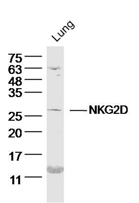
Sample: Lung (Mouse) Lysate at 40 ug
Primary: Anti- NKG2D (SL0938R) at 1/300 dilution
Secondary: IRDye800CW Goat Anti-Rabbit IgG at 1/20000 dilution
Predicted band size: 25 kD
Observed band size: 26 kD
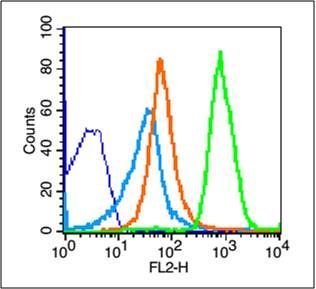
Blank control (blue line): Mouse thymus cells (blue).
Primary Antibody (green line): Rabbit Anti-NKG2D antibody (SL0938R)
Dilution: 1μg /10^6 cells;
Isotype Control Antibody (orange line): Rabbit IgG .
Secondary Antibody (white blue line): Goat anti-rabbit IgG-PE
Dilution: 1μg /test.
Protocol
The cells were fixed with 70% methanol (Overnight at 4℃) . Cells stained with Primary Antibody for 30 min at room temperature. The cells were then incubated in 1 X PBS/2%BSA/10% goat serum to block non-specific protein-protein interactions followed by the antibody for 15 min at room temperature. The secondary antibody used for 40 min at room temperature. Acquisition of 20,000 events was performed.
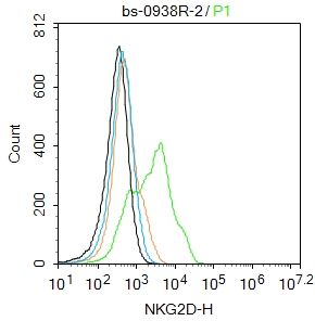
Blank control:SW96.
Primary Antibody (green line): Rabbit Anti-NKG2D antibody (SL0938R)
Dilution: 2ug/Test;
Secondary Antibody : Goat anti-rabbit IgG-FITC
Dilution: 0.5ug/Test.
Protocol
The cells were incubated in 5%BSA to block non-specific protein-protein interactions for 30 min at room temperature .Cells stained with Primary Antibody for 30 min at room temperature. The secondary antibody used for 40 min at room temperature. Acquisition of 20,000 events was performed.
|
|
|