Mitogen-activated protein kinase 4 is a member of the mitogen-activated protein kinase family. Tyrosine kinase growth factor receptors activate mitogen-activated protein kinases which then translocate into the nucleus where it phosphorylates nuclear targets. [provided by RefSeq, Jul 2008]
Function:
Atypical MAPK protein. Phosphorylates microtubule-associated protein 2 (MAP2) and MAPKAPK5. The precise role of the complex formed with MAPKAPK5 is still unclear, but the complex follows a complex set of phosphorylation events: upon interaction with atypical MAPKAPK5, ERK4/MAPK4 is phosphorylated at Ser-186 and then mediates phosphorylation and activation of MAPKAPK5, which in turn phosphorylates ERK4/MAPK4. May promote entry in the cell cycle (By similarity).
Subunit:
Homodimer. Heterodimer with ERK3/MAPK6. Interacts with (via FRIEDE motif) MAPKAPK5.
Subcellular Location:
Cytoplasm. Nucleus. Note=Translocates to the cytoplasm following interaction with MAPKAPK5.
Tissue Specificity:
High expression in heart and brain.
Post-translational modifications:
Phosphorylated at Ser-186 by PAK1, PAK2 and PAK3 resulting in catalytic activation. Phosphorylated by MAPKAPK5 at other sites.
Similarity:
Belongs to the protein kinase superfamily. CMGC Ser/Thr protein kinase family. MAP kinase subfamily.
Contains 1 protein kinase domain.
SWISS:
P31152
Gene ID:
5596
Database links:
Entrez Gene: 5596 Human
Entrez Gene: 225724 Mouse
Entrez Gene: 54268 Rat
Omim: 176949 Human
SwissProt: P31152 Human
SwissProt: Q6P5G0 Mouse
SwissProt: Q63454 Rat
Unigene: 433728 Human
Unigene: 254517 Mouse
Unigene: 146899 Rat
MAP kinase 4(MAPK4)也是MAPK家族成员,具有丝/苏氨酸激酶活性。参与细胞生长、增殖、分化、死亡及细胞间的功能同步等多种生理过程,能够被诸如细胞因子、生长因子、神经递质、激素、细胞应急以及细胞粘附等各种刺激因素激活为信号传导途中重要的蛋白激酶,MAPK家族成员之间有很高的同源性。
| Picture |
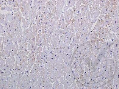
Independently Validated Antibody, image provided by Science Direct, badge number 029727:Formalin-fixed and paraffin embedded human heart labeled with Rabbit Anti-ERK4 Polyclonal Antibody, Unconjugated (SL1319R) at 1:250 overnight at 4C followed by conjugation to the secondary antibody and DAB staining
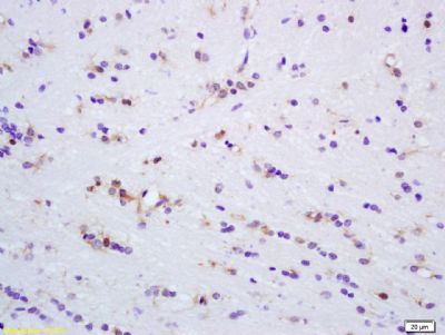
Tissue/cell: rat brain tissue; 4% Paraformaldehyde-fixed and paraffin-embedded;
Antigen retrieval: citrate buffer ( 0.01M, pH 6.0 ), Boiling bathing for 15min; Block endogenous peroxidase by 3% Hydrogen peroxide for 30min; Blocking buffer (normal goat serum,SLC0005) at 37℃ for 20 min;
Incubation: Anti-MAPK4 Polyclonal Antibody, Unconjugated(SL1319R) 1:200, overnight at 4°C, followed by conjugation to the secondary antibody(SP-0023) and DAB(SLC0010) staining
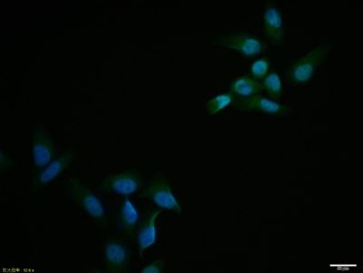
Hela cell; 4% Paraformaldehyde-fixed; Triton X-100 at room temperature for 20 min; Blocking buffer (normal goat serum, SLC0005) at 37°C for 20 min; Antibody incubation with (MAPK4) polyclonal Antibody, Unconjugated (SL1319R) 1:100, 90 minutes at 37°C; followed by a conjugated Goat Anti-Rabbit IgG antibody at 37°C for 90 minutes, DAPI (blue, C02-04002) was used to stain the cell nuclei.
Blank control:Hela.
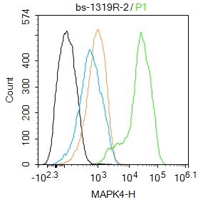
Primary Antibody (green line): Rabbit Anti-MAPK4 antibody (SL1319R)
Dilution: 2ug/Test;
Secondary Antibody : Goat anti-rabbit IgG-FITC
Dilution: 0.5ug/Test.
Protocol
The cells were fixed with 4% PFA (10min at room temperature)and then permeabilized with 90% ice-cold methanol for 20 min at -20℃.The cells were then incubated in 5%BSA to block non-specific protein-protein interactions for 30 min at room temperature .Cells stained with Primary Antibody for 30 min at room temperature. The secondary antibody used for 40 min at room temperature. Acquisition of 20,000 events was performed.
|
|
|